Arquivo da tag: Psiquiatria

‘Everybody has a breaking point’: how the climate crisis affects our brains (Guardian)

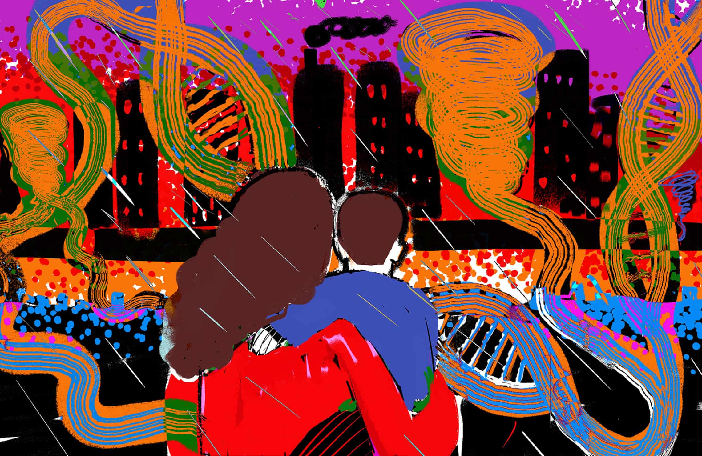
Researchers measuring the effect of Hurricane Sandy on children in utero at the time reported: ‘Our findings are extremely alarming.’ Illustration: Ngadi Smart/The Guardian

Are growing rates of anxiety, depression, ADHD, PTSD, Alzheimer’s and motor neurone disease related to rising temperatures and other extreme environmental changes?

Original article

Clayton Page Aldern

Wed 27 Mar 2024 05.00 GMTShare

In late October 2012, a category 3 hurricane howled into New York City with a force that would etch its name into the annals of history. Superstorm Sandy transformed the city, inflicting more than $60bn in damage, killing dozens, and forcing 6,500 patients to be evacuated from hospitals and nursing homes. Yet in the case of one cognitive neuroscientist, the storm presented, darkly, an opportunity.

Yoko Nomura had found herself at the centre of a natural experiment. Prior to the hurricane’s unexpected visit, Nomura – who teaches in the psychology department at Queens College, CUNY, as well as in the psychiatry department of the Icahn School of Medicine at Mount Sinai – had meticulously assembled a research cohort of hundreds of expectant New York mothers. Her investigation, the Stress in Pregnancy study, had aimed since 2009 to explore the potential imprint of prenatal stress on the unborn. Drawing on the evolving field of epigenetics, Nomura had sought to understand the ways in which environmental stressors could spur changes in gene expression, the likes of which were already known to influence the risk of specific childhood neurobehavioural outcomes such as autism, schizophrenia and attention deficit hyperactivity disorder (ADHD).

The storm, however, lent her research a new, urgent question. A subset of Nomura’s cohort of expectant women had been pregnant during Sandy. She wanted to know if the prenatal stress of living through a hurricane – of experiencing something so uniquely catastrophic – acted differentially on the children these mothers were carrying, relative to those children who were born before or conceived after the storm.

More than a decade later, she has her answer. The conclusions reveal a startling disparity: children who were in utero during Sandy bear an inordinately high risk of psychiatric conditions today. For example, girls who were exposed to Sandy prenatally experienced a 20-fold increase in anxiety and a 30-fold increase in depression later in life compared with girls who were not exposed. Boys had 60-fold and 20-fold increased risks of ADHD and conduct disorder, respectively. Children expressed symptoms of the conditions as early as preschool.

A resident pulls a woman in a canoe down 6th Street as high tide, rain and winds flood local streets on October 29, 2012 in Lindenhurst, New York.
Flooding in Lindenhurst, New York, in October 2012, after Hurricane Sandy struck. Photograph: Bruce Bennett/Getty Images

“Our findings are extremely alarming,” the researchers wrote in a 2022 study summarising their initial results. It is not the type of sentence one usually finds in the otherwise measured discussion sections of academic papers.

Yet Nomura and her colleagues’ research also offers a representative page in a new story of the climate crisis: a story that says a changing climate doesn’t just shape the environment in which we live. Rather, the climate crisis spurs visceral and tangible transformations in our very brains. As the world undergoes dramatic environmental shifts, so too does our neurological landscape. Fossil-fuel-induced changes – from rising temperatures to extreme weather to heightened levels of atmospheric carbon dioxide – are altering our brain health, influencing everything from memory and executive function to language, the formation of identity, and even the structure of the brain. The weight of nature is heavy, and it presses inward.

Evidence comes from a variety of fields. Psychologists and behavioural economists have illustrated the ways in which temperature spikes drive surges in everything from domestic violence to online hate speech. Cognitive neuroscientists have charted the routes by which extreme heat and surging CO2 levels impair decision-making, diminish problem-solving abilities, and short-circuit our capacity to learn. Vectors of brain disease, such as ticks and mosquitoes, are seeing their habitable ranges expand as the world warms. And as researchers like Nomura have shown, you don’t need to go to war to suffer from post-traumatic stress disorder: the violence of a hurricane or wildfire is enough. It appears that, due to epigenetic inheritance, you don’t even need to have been born yet.

When it comes to the health effects of the climate crisis, says Burcin Ikiz, a neuroscientist at the mental-health philanthropy organisation the Baszucki Group, “we know what happens in the cardiovascular system; we know what happens in the respiratory system; we know what happens in the immune system. But there’s almost nothing on neurology and brain health.” Ikiz, like Nomura, is one of a growing cadre of neuroscientists seeking to connect the dots between environmental and neurological wellness.

As a cohesive effort, the field – which we might call climatological neuroepidemiology – is in its infancy. But many of the effects catalogued by such researchers feel intuitive.

Two people trudge along a beach, with the sea behind them, and three folded beach umbrellas standing on the beach. The sky is a dark orange colour and everything in the picture is strongly tinted orange.
Residents evacuate Evia, Greece, in 2021, after wildfires hit the island. Photograph: Bloomberg/Getty Images

Perhaps you’ve noticed that when the weather gets a bit muggier, your thinking does the same. That’s no coincidence; it’s a nearly universal phenomenon. During a summer 2016 heatwave in Boston, Harvard epidemiologists showed that college students living in dorms without air conditioning performed standard cognitive tests more slowly than those living with it. In January of this year, Chinese economists noted that students who took mathematics tests on days above 32C looked as if they had lost the equivalent of a quarter of a year of education, relative to test days in the range 22–24C. Researchers estimate that the disparate effects of hot school days – disproportionately felt in poorer school districts without access to air conditioning and home to higher concentrations of non-white students – account for something on the order of 5% of the racial achievement gap in the US.

Cognitive performance is the tip of the melting iceberg. You may have also noticed, for example, your own feelings of aggression on hotter days. You and everyone else – and animals, too. Black widow spiders tend more quickly toward sibling cannibalism in the heat. Rhesus monkeys start more fights with one another. Baseball pitchers are more likely to intentionally hit batters with their pitches as temperatures rise. US Postal Service workers experience roughly 5% more incidents of harassment and discrimination on days above 32C, relative to temperate days.

Neuroscientists point to a variety of routes through which extreme heat can act on behaviour. In 2015, for example, Korean researchers found that heat stress triggers inflammation in the hippocampus of mice, a brain region essential for memory storage. Extreme heat also diminishes neuronal communication in zebrafish, a model organism regularly studied by scientists interested in brain function. In human beings, functional connections between brain areas appear more randomised at higher temperatures. In other words, heat limits the degree to which brain activity appears coordinated. On the aggression front, Finnish researchers noted in 2017 that high temperatures appear to suppress serotonin function, more so among people who had committed violent crimes. For these people, blood levels of a serotonin transporter protein, highly correlated with outside temperatures, could account for nearly 40% of the fluctuations in the country’s rate of violent crime.

Illustration of a person sweating in an extreme heat scenario
Prolonged exposure to heat can activate a multitude of biochemical pathways associated with Alzheimer’s and Parkinson’s. Illustration: Ngadi Smart/The Guardian

“We’re not thinking about any of this,” says Ikiz. “We’re not getting our healthcare systems ready. We’re not doing anything in terms of prevention or protections.”

Ikiz is particularly concerned with the neurodegenerative effects of the climate crisis. In part, that’s because prolonged exposure to heat in its own right – including an increase of a single degree centigrade – can activate a multitude of biochemical pathways associated with neurodegenerative diseases such as Alzheimer’s and Parkinson’s. Air pollution does the same thing. (In rats, such effects are seen after exposure to extreme heat for a mere 15 minutes a day for one week.) Thus, with continued burning of fossil fuels, whether through direct or indirect effects, comes more dementia. Researchers have already illustrated the manners in which dementia-related hospitalisations rise with temperature. Warmer weather worsens the symptoms of neurodegeneration as well.

Prior to her move to philanthropy, Ikiz’s neuroscience research largely focused on the mechanisms underlying the neurodegenerative disease amyotrophic lateral sclerosis (ALS, also known as Lou Gehrig’s disease or motor neurone disease). Today, she points to research suggesting that blue-green algae, blooming with ever-increasing frequency under a changing global climate, releases a potent neurotoxin that offers one of the most compelling causal explanations for the incidence of non-genetic ALS. Epidemiologists have, for example, identified clusters of ALS cases downwind of freshwater lakes prone to blue-green algae blooms.

A woman pushing a shopping trolley grabs the last water bottles from a long empty shelf in a supermarket.
A supermarket in Long Beach is stripped of water bottles in preparation for Hurricane Sandy. Photograph: Mike Stobe/Getty Images

It’s this flavour of research that worries her the most. Children constitute one of the populations most vulnerable to these risk factors, since such exposures appear to compound cumulatively over one’s life, and neurodegenerative diseases tend to manifest in the later years. “It doesn’t happen acutely,” says Ikiz. “Years pass, and then people get these diseases. That’s actually what really scares me about this whole thing. We are seeing air pollution exposure from wildfires. We’re seeing extreme heat. We’re seeing neurotoxin exposure. We’re in an experiment ourselves, with the brain chronically exposed to multiple toxins.”

Other scientists who have taken note of these chronic exposures resort to similarly dramatic language as that of Nomura and Ikiz. “Hallmarks of Alzheimer disease are evolving relentlessly in metropolitan Mexico City infants, children and young adults,” is part of the title of a recent paper spearheaded by Dr Lilian Calderón-Garcidueñas, a toxicologist who directs the University of Montana’s environmental neuroprevention laboratory. The researchers investigated the contributions of urban air pollution and ozone to biomarkers of neurodegeneration and found physical hallmarks of Alzheimer’s in 202 of the 203 brains they examined, from residents aged 11 months to 40 years old. “Alzheimer’s disease starting in the brainstem of young children and affecting 99.5% of young urbanites is a serious health crisis,” Calderón-Garcidueñas and her colleagues wrote. Indeed.

A flooded Scottish street, with cars standing in water, their wheels just breaking the surface. A row of houses in the background with one shop called The Pet Shop.
Flooding in Stonehaven, Aberdeenshire, in 2020. Photograph: Martin Anderson/PA

Such neurodevelopmental challenges – the effects of environmental degradation on the developing and infant brain – are particularly large, given the climate prognosis. Rat pups exposed in utero to 40C heat miss brain developmental milestones. Heat exposure during neurodevelopment in zebrafish magnifies the toxic effects of lead exposure. In people, early pregnancy exposure to extreme heat is associated with a higher risk of children developing neuropsychiatric conditions such as schizophrenia and anorexia. It is also probable that the ALS-causing neurotoxin can travel in the air.

Of course, these exposures only matter if you make it to an age in which neural rot has a chance to manifest. Neurodegenerative disease mostly makes itself known in middle-aged and elderly people. But, on the other hand, the brain-eating amoeba likely to spread as a result of the climate crisis – which is 97% fatal and will kill someone in a week – mostly infects children who swim in lakes. As children do.

A coordinated effort to fully understand and appreciate the neurological costs of the climate crisis does not yet exist. Ikiz is seeking to rectify this. In spring 2024, she will convene the first meeting of a team of neurologists, neuroscientists and planetary scientists, under the banner of the International Neuro Climate Working Group.

Mexico City landscape engulfed in smog.
Smog hits Mexico City. Photograph: E_Rojas/Getty Images/iStockphoto

The goal of the working group (which, full disclosure, I have been invited to join) is to wrap a collective head around the problem and seek to recommend treatment practices and policy recommendations accordingly, before society finds itself in the midst of overlapping epidemics. The number of people living with Alzheimer’s is expected to triple by 2050, says Ikiz – and that’s without taking the climate crisis into account. “That scares me,” she says. “Because in 2050, we’ll be like: ‘Ah, this is awful. Let’s try to do something.’ But it will be too late for a lot of people.

“I think that’s why it’s really important right now, as evidence is building, as we’re understanding more, to be speaking and raising awareness on these issues,” she says. “Because we don’t want to come to that point of irreversible damage.”

For neuroscientists considering the climate problem, avoiding that point of no return implies investing in resilience research today. But this is not a story of climate anxiety and mental fortitude. “I’m not talking about psychological resilience,” says Nomura. “I’m talking about biological resilience.”

A research agenda for climatological neuroepidemiology would probably bridge multiple fields and scales of analysis. It would merge insights from neurology, neurochemistry, environmental science, cognitive neuroscience and behavioural economics – from molecular dynamics to the individual brain to whole ecosystems. Nomura, for example, wants to understand how external environmental pressures influence brain health and cognitive development; who is most vulnerable to these pressures and when; and which preventive strategies might bolster neurological resilience against climate-induced stressors. Others want to price these stressors, so policymakers can readily integrate them into climate-action cost-benefit analyses.

Wrecked houses along a beach.
Storm devastation in Seaside Heights, New Jersey. Photograph: Mike Groll/AP

For Nomura, it all comes back to stress. Under the right conditions, prenatal exposure to stress can be protective, she says. “It’s like an inoculation, right? You’re artificially exposed to something in utero and you become better at handling it – as long as it is not overwhelmingly toxic.” Stress in pregnancy, in moderation, can perhaps help immunise the foetus against the most deleterious effects of stress later in life. “But everybody has a breaking point,” she says.

Identifying these breaking points is a core challenge of Nomura’s work. And it’s a particularly thorny challenge, in that as a matter of both research ethics and atmospheric physics, she and her colleagues can’t just gin up a hurricane and selectively expose expecting mothers to it. “Human research in this field is limited in a way. We cannot run the gold standard of randomised clinical trials,” she says. “We cannot do it. So we have to take advantage of this horrible natural disaster.”

Recently, Nomura and her colleagues have begun to turn their attention to the developmental effects of heat. They will apply similar methods to those they applied to understanding the effects of Hurricane Sandy – establishing natural cohorts and charting the developmental trajectories in which they’re interested.

The work necessarily proceeds slowly, in part because human research is further complicated by the fact that it takes people longer than animals to develop. Rats zoom through infancy and are sexually mature by about six weeks, whereas for humans it takes more than a decade. “That’s a reason this longitudinal study is really important – and a reason why we cannot just get started on the question right now,” says Nomura. “You cannot buy 10 years’ time. You cannot buy 12 years’ time.” You must wait. And so she waits, and she measures, as the waves continue to crash.

Clayton Page Aldern’s book The Weight of Nature, on the effects of climate change on brain health, is published by Allen Lane on 4 April.

How to mend your broken pandemic brain (MIT Technology Review)

technologyreview.com

Life under covid has messed with our brains. Luckily, they were designed to bounce back.

Dana Smith – July 16, 2021


Orgies are back. Or at least that’s what advertisers want you to believe. One commercial for chewing gum—whose sales tanked during 2020 because who cares what your breath smells like when you’re wearing a mask—depicts the end of the pandemic as a raucous free-for-all with people embracing in the streets and making out in parks. 

The reality is a little different. Americans are slowly coming out of the pandemic, but as they reemerge, there’s still a lot of trauma to process. It’s not just our families, our communities, and our jobs that have changed; our brains have changed too. We’re not the same people we were 18 months ago. 

During the winter of 2020, more than 40% of Americans reported symptoms of anxiety or depression, double the rate of the previous year. That number dropped to 30% in June 2021 as vaccinations rose and covid-19 cases fell, but that still leaves nearly one in three Americans struggling with their mental health. In addition to diagnosable symptoms, plenty of people reported experiencing pandemic brain fog, including forgetfulness, difficulty concentrating, and general fuzziness. 

Now the question is, can our brains change back? And how can we help them do that?

How stress affects the brain

Every experience changes your brain, either helping you to gain new synapses—the connections between brain cells—or causing you to lose them. This is known as neuroplasticity, and it’s how our brains develop through childhood and adolescence. Neuroplasticity is how we continue to learn and create memories in adulthood, too, although our brains become less flexible as we get older. The process is vital for learning, memory, and general healthy brain function.

But many experiences also cause the brain to lose cells and connections that you wanted or needed to keep. For instance, stress—something almost everyone experienced during the pandemic—can not only destroy existing synapses but also inhibit the growth of new ones. 

One way stress does this is by triggering the release of hormones called glucocorticoids, most notably cortisol. In small doses, glucocorticoids help the brain and body respond to a stressor (think: fight or flight) by changing heart rate, respiration, inflammation, and more to increase one’s odds of survival. Once the stressor is gone, the hormone levels recede. With chronic stress, however, the stressor never goes away, and the brain remains flooded with the chemicals. In the long term, elevated levels of glucocorticoids can cause changes that may lead to depression, anxiety, forgetfulness, and inattention. 

Scientists haven’t been able to directly study these types of physical brain changes during the pandemic, but they can make inferences from the many mental health surveys conducted over the last 18 months and what they know about stress and the brain from years of previous research.

For example, one study showed that people who experienced financial stressors, like a job loss or economic insecurity, during the pandemic were more likely to develop depression. One of the brain areas hardest hit by chronic stress is the hippocampus, which is important for both memory and mood. These financial stressors would have flooded the hippocampus with glucocorticoids for months, damaging cells, destroying synapses, and ultimately shrinking the region. A smaller hippocampus is one of the hallmarks of depression. 

Chronic stress can also alter the prefrontal cortex, the brain’s executive control center, and the amygdala, the fear and anxiety hub. Too many glucocorticoids for too long can impair the connections both within the prefrontal cortex and between it and the amygdala. As a result, the prefrontal cortex loses its ability to control the amygdala, leaving the fear and anxiety center to run unchecked. This pattern of brain activity (too much action in the amygdala and not enough communication with the prefrontal cortex) is common in people who have post-traumatic stress disorder (PTSD), another condition that spiked during the pandemic, particularly among frontline health-care workers.

The social isolation brought on by the pandemic was also likely detrimental to the brain’s structure and function. Loneliness has been linked to reduced volume in the hippocampus and amygdala, as well as decreased connectivity in the prefrontal cortex. Perhaps unsurprisingly, people who lived alone during the pandemic experienced higher rates of depression and anxiety.

Finally, damage to these brain areas affects people not only emotionally but cognitively as well. Many psychologists have attributed pandemic brain fog to chronic stress’s impact on the prefrontal cortex, where it can impair concentration and working memory.

Reversal time

So that’s the bad news. The pandemic hit our brains hard. These negative changes ultimately come down to a stress-induced decrease in neuroplasticity—a loss of cells and synapses instead of the growth of new ones. But don’t despair; there’s some good news. For many people, the brain can spontaneously recover its plasticity once the stress goes away. If life begins to return to normal, so might our brains.

“In a lot of cases, the changes that occur with chronic stress actually abate over time,” says James Herman, a professor of psychiatry and behavioral neuroscience at the University of Cincinnati. “At the level of the brain, you can see a reversal of a lot of these negative effects.” 

“If you create for yourself a more enriched environment where you have more possible inputs and interactions and stimuli, then [your brain] will respond to that.”

Rebecca Price, associate professor of psychiatry and psychology at the University of Pittsburgh

In other words, as your routine returns to its pre-pandemic state, your brain should too. The stress hormones will recede as vaccinations continue and the anxiety about dying from a new virus (or killing someone else) subsides. And as you venture out into the world again, all the little things that used to make you happy or challenged you in a good way will do so again, helping your brain to repair the lost connections that those behaviors had once built. For example, just as social isolation is bad for the brain, social interaction is especially good for it. People with larger social networks have more volume and connections in the prefrontal cortexamygdala, and other brain regions. 

Even if you don’t feel like socializing again just yet, maybe push yourself a little anyway. Don’t do anything that feels unsafe, but there is an aspect of “fake it till you make it” in treating some mental illness. In clinical speak, it’s called behavioral activation, which emphasizes getting out and doing things even if you don’t want to. At first, you might not experience the same feelings of joy or fun you used to get from going to a bar or a backyard barbecue, but if you stick with it, these activities will often start to feel easier and can help lift feelings of depression.

Rebecca Price, an associate professor of psychiatry and psychology at the University of Pittsburgh, says behavioral activation might work by enriching your environment, which scientists know leads to the growth of new brain cells, at least in animal models. “Your brain is going to react to the environment that you present to it, so if you are in a deprived, not-enriched environment because you’ve been stuck at home alone, that will probably cause some decreases in the pathways that are available,” she says. “If you create for yourself a more enriched environment where you have more possible inputs and interactions and stimuli, then [your brain] will respond to that.” So get off your couch and go check out a museum, a botanical garden, or an outdoor concert. Your brain will thank you.

Exercise can help too. Chronic stress depletes levels of an important chemical called brain-derived neurotrophic factor (BDNF), which helps promote neuroplasticity. Without BDNF, the brain is less able to repair or replace the cells and connections that are lost to chronic stress. Exercise increases levels of BDNF, especially in the hippocampus and prefrontal cortex, which at least partially explains why exercise can boost both cognition and mood. 

Not only does BDNF help new synapses grow, but it may help produce new neurons in the hippocampus, too. For decades, scientists thought that neurogenesis in humans stopped after adolescence, but recent research has shown signs of neuron growth well into old age (though the issue is still hotly contested). Regardless of whether it works through neurogenesis or not, exercise has been shown time and again to improve people’s mood, attention, and cognition; some therapists even prescribe it to treat depression and anxiety. Time to get out there and start sweating.

Turn to treatment

There’s a lot of variation in how people’s brains recover from stress and trauma, and not everyone will bounce back from the pandemic so easily.

“Some people just seem to be more vulnerable to getting into a chronic state where they get stuck in something like depression or anxiety,” says Price. In these situations, therapy or medication might be required.

Some scientists now think that psychotherapy for depression and anxiety works at least in part by changing brain activity, and that getting the brain to fire in new patterns is a first step to getting it to wire in new patterns. A review paper that assessed psychotherapy for different anxiety disorders found that the treatment was most effective in people who displayed more activity in the prefrontal cortex after several weeks of therapy than they did beforehand—particularly when the area was exerting control over the brain’s fear center. 

Other researchers are trying to change people’s brain activity using video games. Adam Gazzaley, a professor of neurology at the University of California, San Francisco, developed the first brain-training game to receive FDA approval for its ability to treat ADHD in kids. The game has also been shown to improve attention span in adults. What’s more, EEG studies revealed greater functional connectivity involving the prefrontal cortex, suggesting a boost in neuroplasticity in the region.

Now Gazzaley wants to use the game to treat people with pandemic brain fog. “We think in terms of covid recovery there’s an incredible opportunity here,” he says. “I believe that attention as a system can help across the breadth of [mental health] conditions and symptoms that people are suffering, especially due to covid.”

While the effects of brain-training games on mental health and neuroplasticity are still up for debate, there’s abundant evidence for the benefits of psychoactive medications. In 1996, psychiatrist Yvette Sheline, now a professor at the University of Pennsylvania, was the first to show that people with depression had significantly smaller hippocampi than non-depressed people, and that the size of that brain region was related to how long and how severely they had been depressed. Seven years later, she found that if people with depression took antidepressants, they had less volume loss in the region.

That discovery shifted many researchers’ perspectives on how traditional antidepressants, particularly selective serotonin reuptake inhibitors (SSRIs), help people with depression and anxiety. As their name suggests, SSRIs target the neurochemical serotonin, increasing its levels in synapses. Serotonin is involved in several basic bodily functions, including digestion and sleep. It also helps to regulate mood, and scientists long assumed that was how the drugs worked as antidepressants. However, recent research suggests that SSRIs may also have a neuroplastic effect by boosting BDNF, especially in the hippocampus, which could help restore healthy brain function in the area. One of the newest antidepressants approved in the US, ketamine, also appears to increase BDNF levels and promote synapse growth in the brain, providing additional support for the neuroplasticity theory. 

The next frontier in pharmaceutical research for mental illness involves experimental psychedelics like MDMA and psilocybin, the active ingredient in hallucinogenic mushrooms. Some researchers think that these drugs also enhance plasticity in the brain and, when paired with psychotherapy, can be a powerful treatment.

Not all the changes to our brains from the past year are negative. Neuroscientist David Eagleman, author of the book Livewired: The Inside Story of the Ever-Changing Brain, says that some of those changes may actually have been beneficial. By forcing us out of our ruts and changing our routines, the pandemic may have caused our brains to stretch and grow in new ways.

“This past 14 months have been full of tons of stress, anxiety, depression—they’ve been really hard on everybody,” Eagleman says. “The tiny silver lining is from the point of view of brain plasticity, because we have challenged our brains to do new things and find new ways of doing things. If we hadn’t experienced 2020, we’d still have an old internal model of the world, and we wouldn’t have pushed our brains to make the changes they’ve already made. From a neuroscience point of view, this is most important thing you can do—constantly challenge it, build new pathways, find new ways of seeing the world.”


How to help your brain help itself

While everyone’s brain is different, try these activities to give your brain the best chance of recovering from the pandemic.

  1. Get out and socialize. People with larger social networks have more volume and connectivity in the prefrontal cortexamygdala, and other brain regions.
  2. Try working out. Exercise increases levels of a protein called BDNF that helps promote neuroplasticity and may even contribute to the growth of new neurons.
  3. Talk to a therapist. Therapy can help you view yourself from a different perspective, and changing your thought patterns can change your brain patterns.
  4. Enrich your environment. Get out of your pandemic rut and stimulate your brain with a trip to the museum, a botanical garden, or an outdoor concert.
  5. Take some drugs—but make sure they’re prescribed! Both classic antidepressant drugs, such as SSRIs, and more experimental ones like ketamine and psychedelics are thought to work in part by boosting neuroplasticity.
  6. Strengthen your prefrontal cortex by exercising your self-control. If you don’t have access to an (FDA-approved) attention-boosting video game, meditation can have a similar benefit. 

4 efeitos do racismo no cérebro e no corpo de crianças, segundo Harvard (BBC)

Paula Adamo Idoeta

Da BBC News Brasil em São Paulo

9 dezembro 2020, 06:01 -03

Criança com a mãe
Viver o racismo, direta ou indiretamente, tem efeitos de longo prazo sobre desenvolvimento, comportamento, saúde física e mental

Episódios diários de racismo, desde ser alvo de preconceito até assistir a casos de violência sofridos por outras pessoas da mesma raça, têm um efeito às vezes “invisível”, mas duradouro e cruel sobre a saúde, o corpo e o cérebro de crianças.

A conclusão é do Centro de Desenvolvimento Infantil da Universidade de Harvard, que compilou estudos documentando como a vivência cotidiana do racismo estrutural, de suas formas mais escancaradas às mais sutis ou ao acesso pior a serviços públicos, impacta “o aprendizado, o comportamento, a saúde física e mental” infantil.

No longo prazo, isso resulta em custos bilionários adicionais em saúde, na perpetuação das disparidades raciais e em mais dificuldades para grande parcela da população em atingir seu pleno potencial humano e capacidade produtiva.

Embora os estudos sejam dos EUA, dados estatísticos — além do fato de o Brasil também ter histórico de escravidão e desigualdade — permitem traçar paralelos entre os dois cenários.

Aqui, casos recentes de violência contra pessoas negras incluem o de Beto Freitas, espancado até a morte dentro de um supermercado Carrefour em Porto Alegre em 20 de novembro, e o das primas Emilly, 4, e Rebeca, 7, mortas por disparos de balas enquanto brincavam na porta de casa, em Duque de Caxias em 4 de dezembro.

No Brasil, 54% da população é negra, percentual que é de 13% na população dos EUA.

A seguir, quatro impactos do ciclo vicioso do racismo, segundo o documento de Harvard. Para discutir as particularidades disso no Brasil, a reportagem entrevistou a psicóloga Cristiane Ribeiro, autora de um estudo recente sobre como a população negra lida com o sofrimento físico e mental, que foi tema de sua dissertação de mestrado pelo Programa de Pós-graduação em Promoção da Saúde e Prevenção da Violência da UFMG.

1. Corpo em estado de alerta constante

O racismo e a violência dentro da comunidade (e a ausência de apoio para lidar com isso) estão entre o que Harvard chama de “experiências adversas na infância”. Passar constantemente por essas experiências faz com que o cérebro se mantenha em estado constante de alerta, provocando o chamado “estresse tóxico”.

“Anos de estudos científicos mostram que, quando os sistemas de estresse das crianças ficam ativados em alto nível por longo período de tempo, há um desgaste significativo nos seus cérebros em desenvolvimento e outros sistemas biológicos”, diz o Centro de Desenvolvimento Infantil da universidade.

Na prática, áreas do cérebro dedicadas à resposta ao medo, à ansiedade e a reações impulsivas podem produzir um excesso de conexões neurais, ao mesmo tempo em que áreas cerebrais dedicadas à racionalização, ao planejamento e ao controle de comportamento vão produzir menos conexões neurais.

Protesto pela morte de Beto Freitas, em Porto Alegre, 20 de novembro
Protesto pela morte de Beto Freitas, em Porto Alegre, 20 de novembro; assistir cenas de violência contra pessoas da mesma raça tem efeito traumático – é o chamado ‘racismo indireto’

“Isso pode ter efeito de longo prazo no aprendizado, comportamento, saúde física e mental”, prossegue o centro. “Um crescente corpo de evidências das ciências biológicas e sociais conecta esse conceito de desgaste (do cérebro) ao racismo. Essas pesquisas sugerem que ter de lidar constantemente com o racismo sistêmico e a discriminação cotidiana é um ativador potente da resposta de estresse.”

“Embora possam ser invisíveis para quem não passa por isso, não há dúvidas de que o racismo sistêmico e a discriminação interpessoal podem levar à ativação crônica do estresse, impondo adversidades significativas nas famílias que cuidam de crianças pequenas”, conclui o documento de Harvard.

2. Mais chance de doenças crônicas ao longo da vida

Essa exposição ao estresse tóxico é um dos fatores que ajudam a explicar diferenças raciais na incidência de doenças crônicas, prossegue o centro de Harvard:

“As evidências são enormes: pessoas negras, indígenas e de outras raças nos EUA têm, em média, mais problemas crônicos de saúde e vidas mais curtas do que as pessoas brancas, em todos os níveis de renda.”

Alguns dados apontam para situação semelhante no Brasil. Homens e mulheres negros têm, historicamente, incidência maior de diabetes — 9% mais prevalente em negros do que em brancos; 50% mais prevalente em negras do que em brancas, segundo o Ministério da Saúde — e pressão alta, por exemplo.

Os números mais marcantes, porém, são os de violência armada, como a que vitimou as meninas Emilly e Rebeca. O Atlas da Violência aponta que negros foram 75,7% das vítimas de homicídio no Brasil em 2018.

A taxa de homicídios de brasileiros negros é de 37,8 para cada 100 mil habitantes, contra 13,9 de não negros.

Há, ainda, uma incidência possivelmente maior de problemas de saúde mental: de cada dez suicídios em adolescentes em 2016, seis foram de jovens negros e quatro de brancos, segundo pesquisa do Ministério da Saúde publicada no ano passado.

“O adoecimento (pela vivência do racismo) é constante, e vemos nos dados escancarados, como os da violência, mas também na depressão, no adoecimento psíquico e nos altos números de suicídio”, afirma a psicóloga Cristiane Ribeiro.

Protesto pela morte de Beto Freitas
“Embora possam ser invisíveis para quem não passa por isso, não há dúvidas de que o racismo sistêmico e a discriminação interpessoal podem levar à ativação crônica do estresse, impondo adversidades significativas nas famílias que cuidam de crianças pequenas”, diz o documento de Harvard

“E por que essa é violência é tão marcante entre pessoas negras? Porque aprendemos que nosso semelhante é o pior possível e o quanto mais longe estivermos dele, melhor. A criança materializa isso de alguma forma. Temos estatísticas de que crianças negras são menos abraçadas na educação infantil, recebem menos afeto dos professores. (Algumas) ouvem desde cedo ‘esse menino não aprende mesmo, é burro’ ou ‘nasceu pra ser bandido'”, prossegue Ribeiro.

Embora muitos conseguem superar essa narrativa, outros têm sua vida marcada por ela, diz Ribeiro. “Trabalhei durante muito tempo no sistema socioeducativo (com jovens infratores), e essas sentenças são muito recorrentes: o menino que escuta desde pequeno que ‘não vai ser nada na vida’. São trajetórias sentenciadas.”

3. Disparidades na saúde e na educação

Os problemas descritos acima são potencializados pelo menor acesso aos serviços públicos de saúde, aponta Harvard.

“Pessoas de cor recebem tratamento desigual quando interagem em sistemas como o de saúde e educação, além de terem menos acesso a educação e serviços de saúde de alta qualidade, a oportunidades econômicas e a caminhos para o acúmulo de riqueza”, diz o documento do Centro de Desenvolvimento infantil.

“Tudo isso reflete formas como o legado do racismo estrutural nos EUA desproporcionalmente enfraquece a saúde e o desenvolvimento de crianças de cor.”

Mais uma vez, os números brasileiros apontam para um quadro parecido. Segundo levantamento do Ministério da Saúde, 67% do público do SUS (Sistema Único de Saúde) é negro. No entanto, a população negra realiza proporcionalmente menos consultas médicas e atendimentos de pré-natal.

E, entre os 10% de pessoas com menor renda no Brasil, 75% delas são pretas ou pardas.

Na educação, as disparidades persistem. Crianças negras de 0 a 3 anos têm percentual menor de matrículas em creches. Na outra ponta do ensino, 53,9% dos jovens declarados negros concluíram o ensino médio até os 19 anos — 20 pontos percentuais a menos que a taxa de jovens brancos, apontam dados de 2018 do movimento Todos Pela Educação.

Familiares das meninas Emilly e Rebecca, mortas a tiros,em encontro com o governador em exercício do Rio, Claudio Castro
Familiares das meninas Emilly e Rebecca, mortas a tiros,em encontro com o governador em exercício do Rio, Claudio Castro; Atlas da Violência aponta que negros foram 75,7% das vítimas de homicídio no Brasil em 2018

4. Cuidadores mais fragilizados e ‘racismo indireto’

Os efeitos do estresse não se limitam às crianças: se estendem também aos pais e responsáveis por elas — e, como em um efeito bumerangue, voltam a afetar as crianças indiretamente.

“Múltiplos estudos documentaram como os estresses da discriminação no dia a dia em pais e outros cuidadores, como ser associado a estereótipos negativos, têm efeitos nocivos no comportamento desses adultos e em sua saúde mental”, prossegue o Centro de Desenvolvimento Infantil.

Um dos estudos usados para embasar essa conclusão é uma revisão de dezenas de pesquisas clínicas feita em 2018, que aborda o que os pesquisadores chamam de “exposição indireta ao racismo”: mesmo quando as crianças não são alvo direto de ofensas ou violência racista, podem ficar traumatizadas ao testemunhar ou escutar sobre eventos que tenham afetado pessoas próximas a elas.

“Especialmente para crianças de minorias (raciais), a exposição frequente ao racismo indireto pode forçá-las a dar sentido cognitivamente a um mundo que sistematicamente as desvaloriza e marginaliza”, concluem os pesquisadores.

O estudo identificou, como efeito desse “racismo indireto”, impactos tanto em cuidadores (que tinham autoestima mais fragilizada) como nas crianças, que nasciam de mais partos prematuros, com menor peso ao nascer e mais chances de adoecer ao longo da vida ou de desenvolver depressão.

Na infância, diz a psicóloga Cristiane Ribeiro, é quando começamos a construir nossa capacidade de acreditar no próprio potencial para viver no mundo. No caso da população negra, essa construção é afetada negativamente pelos estereótipos racistas, sejam características físicas ou sociais — como o “cabelo pixaim” ou “serviço de preto”.

Homem penteando cabelo de menina negra
Valorização e representatividade impactam positivamente as crianças e, por consequência, suas famílias

“A gente precisa ter referências mais positivas da população negra como aquela que também é responsável pela constituição social do Brasil. A única representação que a gente tem no livro didático de história é de uma pessoa (escravizada) acorrentada, em uma situação de extrema vulnerabilidade e que está ali porque ‘não se esforçou para não estar'”, diz a pesquisadora.

Mesmo atos “sutis” — como pessoas negras sendo seguidas por seguranças em shopping centers ou recebendo atendimento pior em uma loja qualquer —, que muitas vezes passam despercebidos para observadores brancos, podem ter efeitos devastadores sobre a autoestima, prossegue Ribeiro.

“Isso que a gente costuma chamar de sutileza do racismo não tem nada de sutil na minha perspectiva. Quando alguém grita ‘macaco’ no meio da rua, as pessoas compartilham a indignação. É diferente do olhar (preconceituoso), que só o sujeito viu e só ele percebeu. Mesmo para a militante mais empoderada e ciente de seus direitos — porque é uma luta sem descanso —, tem dias que não tem jeito, esse olhar te destroça. A gente fala muito da força da mulher negra, mas e o direito à fragilidade? será que ser frágil também é um privilégio?”

Como romper o ciclo

“Avanços na ciência apresentam um retrato cada vez mais claro de como a adversidade forte na vida de crianças pequenas pode afetar o desenvolvimento do cérebro e outros sistemas biológicos. Essas perturbações iniciais podem enfraquecer as oportunidades dessas crianças em alcançar seu pleno potencial”, diz o documento de Harvard.

Mas é possível romper esse ciclo, embora lembrando que as formas de combatê-lo são complexas e múltiplas.

Cristiane Ribeiro
“A gente fala muito da força da mulher negra, mas e o direito à fragilidade? será que ser frágil também é um privilégio?”, diz Cristiane Ribeiro

“Precisamos criar novas estratégias para lidar com essas desigualdades que sistematicamente ameaçam a saúde e o bem-estar das crianças pequenas de cor e os adultos que cuidam delas. Isso inclui buscar ativamente e reduzir os preconceitos em nós e nas políticas socioeconômicas, por meio de iniciativas como contratações justas, oferta de crédito, programas de habitação, treinamento antipreconceito e iniciativas de policiamento comunitário”, diz o Centro de Desenvolvimento Infantil de Harvard.

Para Cristiane Ribeiro, passos fundamentais nessa direção envolvem mais representatividade negra e mais discussões sobre o tema dentro das escolas.

“Se tenho uma escola repleta de negros ou pessoas de diferentes orientações sexuais, mas isso não é dito, não é tratado, você tem a mesma segregação que nos outros espaços”, opina.

“Precisamos extinguir a ideia do ‘lápis cor de pele’. Tem tanta cor de pele, porque um lápis rosa a representa? Tem também a criança com cabelo crespo em uma escola onde só são penteados os cabelos lisos. Se a professora der conta de tratar aquele cabelo de uma forma tão afetiva quanto ela trata o cabelo lisinho, ela mudará o mundo daquela criança, inclusive incluindo nessa criança defesa para que ela responda quando seu cabelo for chamado de duro, de feio. E daí ela se olha no espelho e vê beleza, que é um direito que está sendo conquistado muito aos poucos. A chance é de que faça diferença pra família inteira. A criança negra que fala ‘não, mãe, meu cabelo não é feio’ desloca aquele ciclo naquela família, de todas as mulheres alisarem o cabelo. (…) Um olhar afetivo nessa história quebra o ciclo.”

O afeto e a construção de redes de apoio também são apontados por Harvard como formas de aliviar o peso do estresse tóxico e construir resiliência em crianças e famílias.

“É claro que a ciência não consegue lidar com esses desafios sozinha, mas o pensamento informado pela ciência combinado com o conhecimento em mudar sistemas entrincheirados e as experiências vividas pelas famílias que criam seus filhos sob diferentes condições podem ser poderosos catalisadores de estratégias eficientes,” defende o Centro para o Desenvolvimento Infantil.

Como a educação brasileira acentua desigualdade racial e apaga os heróis negros da história do Brasil

Crianças reproduzem racismo? O debate que transformou escola em SP

Why nutritional psychiatry is the future of mental health treatment (The Conversation)

A lack of essential nutrients is known to contribute to the onset of poor mental health in people suffering from anxiety and depression, bipolar disorder, schizophrenia and ADHD. Nutritional psychiatry is a growing discipline that focuses on the use of food and supplements to provide these essential nutrients as part of an integrated or alternative treatment for mental health disorders.

But nutritional approaches for these debilitating conditions are not widely accepted by mainstream medicine. Treatment options tend to be limited to official National Institute for Care Excellence (NICE) guidelines which recommend talking therapies and antidepressants.

Use of antidepressants

Antidepressant use has more than doubled in recent years. In England 64.7m prescriptions were issued for antidepressants in 2016 at a cost of £266.6m. This is an increase of 3.7m on the number of items prescribed in 2015 and more than double than the 31m issued in 2006.

A recent Oxford University study found that antidepressants were more effective in treating depression than placebo. The study was led by Dr Andrea Cipriani who claimed that depression is under treated. Cipriani maintains that antidepressants are effective and a further 1m prescriptions should be issued to people in the UK.

This approach suggests that poor mental health caused by social conditions is viewed as easily treated by simply dispensing drugs. But antidepressants are shunned by people whom they could help because of the social stigma associated with mental ill-health which leads to discrimination and exclusion.

Prescriptions for 64.7m items of antidepressants were dispensed in England in 2016, the highest level recorded by the NHS. Shutterstock

More worrying is the increase in the use of antidepressants by children and young people. In Scotland, 5,572 children under 18 were prescribed antidepressants for anxiety and depression in 2016. This figure has more than doubled since 2009/2010.

But according to British psychopharmacologist Professor David Healy, 29 clinical trials of antidepressant use in young people found no benefits at all. These trials revealed that instead of relieving symptoms of anxiety and depression, antidepressants caused children and young people to feel suicidal.

Healy also challenges their safety and effectiveness in adults. He believes that antidepressants are over-prescribed and that there is little evidence that they are safe for long-term use. Antidepressants are said to create dependency, have unpleasant side effects and cannot be relied upon to always relieve symptoms.

Nutrition and poor mental health

In developed countries such as the UK people eat a greater variety of foodstuffs than ever before – but it doesn’t follow that they are well nourished. In fact, many people do not eat enough nutrients that are essential for good brain health, opting for a diet of heavily processed food containing artificial additives and sugar.

The link between poor mental health and nutritional deficiencies has long been recognised by nutritionists working in the complementary health sector. However, psychiatrists are only now becoming increasingly aware of the benefits of using nutritional approaches to mental health, calling for their peers to support and research this new field of treatment.

It is now known that many mental health conditions are caused by inflammation in the brain which ultimately causes our brain cells to die. This inflammatory response starts in our gut and is associated with a lack of nutrients from our food such as magnesium, omega-3 fatty acids, probiotics, vitamins and minerals that are all essential for the optimum functioning of our bodies.

Recent research has shown that food supplements such as zinc, magnesium, omega 3, and vitamins B and D3 can help improve people’s mood, relieve anxiety and depression and improve the mental capacity of people with Alzheimer’s.

Magnesium is one of most important minerals for optimal health, yet many people are lacking in it. One studyfound that a daily magnesium citrate supplement led to a significant improvement in depression and anxiety, regardless of age, gender or severity of depression. Improvement did not continue when the supplement was stopped.

Omega-3 fatty acids are another nutrient that is critical for the development and function of the central nervous system – and a lack has been associated with low mood, cognitive decline and poor comprehension.

Research has shown that supplements like zinc, magnesium and vitamins B and D can improve the mental capacity of people with Alzheimer’s. Shutterstock

The role of probiotics – the beneficial live bacteria in your digestive system – in improving mental health has also been explored by psychiatrists and nutritionists, who found that taking them daily was associated with a significant reduction in depression and anxiety. Vitamin B complex and zinc are other supplements found to reduce the symptoms of anxiety and depression.

Hope for the future?

These over-the-counter” supplements are widely available in supermarkets, chemists and online health food stores, although the cost and quality may vary. For people who have not responded to prescription drugs or who cannot tolerate the side effects, nutritional intervention can offer hope for the future.

There is currently much debate over the effectiveness of antidepressants. The use of food supplements offer an alternative approach that has the potential to make a significant difference to the mental health of all age groups.

The emerging scientific evidence suggests that there should be a bigger role for nutritional psychiatry in mental health within conventional health services. If the burden of mental ill health is to be reduced, GPs and psychiatrists need to be aware of the connection between food, inflammation and mental illness.

Medical education has traditionally excluded nutritional knowledge and its association with disease. This has led to a situation where very few doctors in the UK have a proper understanding of the importance of nutrition. Nutritional interventions are thought to have little evidence to support their use to prevent or maintain well-being and so are left to dietitians, rather than doctors, to advise on.

But as the evidence mounts up, it is time for medical education to take nutrition seriously so that GPs and psychiatrists of the future know as much about its role in good health as they do about anatomy and physiology. The state of our mental health could depend on it.

Estamos todos doentes? (JC)

JC, 5707, 25 de julho de 2017

Pesquisadora da Unicamp alerta para influência da indústria farmacêutica no crescimento do número de diagnósticos de transtornos mentais

Dados do National Institute of Mental Health (NIMH, 2012) apontam que 46% dos norte-americanos preenchem os critérios de diagnóstico de um transtorno mental. Na Europa essa porcentagem corresponde a 38%. Nos Estados Unidos, o diagnóstico de transtorno bipolar em crianças e adolescentes aumentou 40 vezes, entre 1994 e 2003, e uma entre cinco crianças tem um surto de transtorno mental por ano, de acordo com dados do Centro de Controle de Doenças, daquele país (CDC, 2013).

Há pesquisas que indicam que 10% da população mundial teria algum tipo de transtorno, um número que segundo a médica pediatra, Maria Aparecida Affonso Moysés, da Faculdade de Medicina da Universidade Estadual de Campinas (Unicamp), inviabiliza qualquer esforço de política pública. “Temos que começar a questionar como esses números são construídos. Na verdade, mudanças nos critérios do diagnóstico se tornaram muito frouxos nos últimos anos”, afirmou a médica, em sua conferência na Reunião Anual da SBPC. “É muito difícil qualquer um de nós não se encaixar nos critérios. Os testes que detectam algumas dessas doenças são verdadeiras armadilhas”, alertou.

Ela refutou a ideia de que vivemos uma epidemia de doenças mentais. “O que temos é uma epidemia de diagnósticos de transtornos mentais”, disse. Na primeira vez em que foi publicado pela Associação Americana de Psiquiatria, em 1952, o Manual Diagnóstico e Estatístico de Transtornos Mentais (DSM), tinha 106 categorias de transtornos mentais. Em sua última edição, em 2013, foram listadas 300 categorias. “Alterar as normas para caracterização de um transtorno e criar doenças novas contribuíram para essa epidemia amplamente patrocinada pela indústria farmacêutica”, declarou Moysés. “Antes de vender remédios, o departamento de marketing da indústria de fármacos trabalha para vender doenças”, diz. Déficit de atenção, transtorno de descontrole de humor, transtorno de aprendizagem, depressão, transtorno opositor desafiante, hiperatividade, são algumas dessas doenças fabricadas para vender medicamentos. “Onde está a ciência e ética nesse campo?”, questionou a médica. Segundo ela, esses medicamentos são largamente receitados para crianças e adultos, como se fossem 100% seguros, mas boa parte deles provoca dependência química.

Um exemplo é o metilfenidato, base de uma classe de estimulantes do sistema nervoso central, vendido entre outras, com a marca “Ritalina”. O medicamento age inibindo a receptação de dopamina na sinapse, o que teria como resultado o aumento do nível de concentração. Conforme explicou Moysés, ele é receitado para crianças com Transtorno do Déficit de Atenção com Hiperatividade (TDAH), não pelo seu efeito terapêutico, mas pelas reações adversas. No sistema nervoso central o metilfenidato provoca o efeito “zombie like”, quando a pessoa fica contida em si mesma. “Eu comparo esse fármaco a uma droga da obediência porque o indivíduo perde a capacidade de questionar, de sentir. É um tipo de contenção química. O aumento de concentração, tão propagado, é, na verdade, uma redução do foco da atenção, isto é, a pessoa presta atenção em uma coisa de cada vez”, afirmou. “Não existe uma pílula que nos faça prestar atenção. Para isso, precisamos de bons professores com boas condições de trabalho”.

Na opinião da pesquisadora da Unicamp, vivemos em um projeto de sociedade que estimula e premia comportamentos homogêneos, punindo as singularidades. “Não é à toa que assistimos cortes significativos nos orçamentos da ciência e da educação. Temos que ser iguais porque as diferenças incomodam cada vez mais. Entretanto, neutralizar os sonhadores, os que pensam diferente é um genocídio do futuro”, disse.

O combate ao que a médica chama de patologização da sociedade passa pelos campos da saúde, da educação e por uma revisão das políticas públicas para que elas não sejam submissas ao mercado. Outro setor é o da formação profissional. Nas escolas de medicina, a técnica não pode se sobrepor à ética e ao aspecto humano. Toda avaliação e diagnóstico têm que respeitar saberes, valores, história e a cultura porque “a vida não é mercadoria”, finalizou.

Por Patricia Mariuzzo para o Jornal da Ciência

Médiuns têm perfil diferente daquele apresentado na literatura científica (USP Notícias)

11/05/2005

Estudo com 115 médiuns kardecistas de São Paulo indica que a maioria possui alto nível socioeducacional, perfil que se enquadra no último censo do IBGE. Segundo a pesquisa, eles não apresentam problemas mentais

Na literatura científica, muitas vezes os médiuns (que se comunicam com espíritos) são descritos como pessoas de baixa escolaridade e renda. Sua mediunidade deve ser entendida como um “mecanismo de defesa contra as opressões sociais”, ou como manifestação de algum quadro dissociativo ou psicótico.

No entanto, um estudo realizado pelo psiquiatra Alexander Moreira de Almeida com médiuns espíritas da cidade de São Paulo mostrou um perfil diferente: os médiuns apresentaram um alto nível socioeducacional e uma prevalência de transtornos mentais menor do que a encontrada na população em geral.

Almeida constatou que 46,5% das pessoas tinham curso superior, 76,5% eram mulheres, menos de 3% estavam desempregados, e a idade média era de 48 anos. A maioria era espírita há mais de 16 anos, vieram de famílias não-espíritas e as vivências mediúnicas começaram na infância.

“Esse perfil sociodemográfico se encaixa no último censo do Instituto Brasileiro de Geografia e Estatística (IBGE), que mostra um crescimento da proporção de espíritas conforme aumenta a escolaridade da população”, comenta o psiquiatra, que apresentou sua tese de doutorado à Faculdade de Medicina (FMUSP), com orientação do professor Francisco Lotufo Neto.

Os participantes do estudo atuam em nove centros espíritas kardecistas da Capital, pertencentes à Aliança Espírita Evangélica. O médico aplicou um questionário sóciodemográfico a 115 médiuns antes e depois das sessões espíritas. Eles também responderam a questões referentes à atividade mediúnica. Almeida ainda utilizou os questionários SRQ (Self-Report Psychiatric Screening Questionnaire), que rastreia a presença de transtornos mentais, e o EAS (Escala de Adequação Social), que mostra como a pessoa se relaciona em sociedade.

A partir dos resultados foram selecionados 24 médiuns. Eles foram analisados pelo SCAN (Schedules for Clinical Assessment in Neuropsychiatry), um tipo de entrevista psiquiátrica padrão e pelo DDIS (Dissociative Disorders Interview Schedule), um questionário que detecta transtornos dissociativos (quando uma parte da mente funciona de forma independente). “É nessa categoria que os transes mediúnicos são habitualmente encaixados”, explica o médico.

“Os médiuns apresentaram, em média, quatro sintomas de primeira ordem para diagnóstico de esquizofrenia, mas a presença desses sintomas não indicou a existência de nenhuma doença mental”

Transes X esquizofrenia

A escala DDIS investiga a presença de 11 sintomas de primeira ordem para o diagnóstico de esquizofrenia – vozes dialogando na sua cabeça, vozes comentando as suas ações, ter suas ações produzidas ou controladas por alguém ou algo fora de você, entre outros. “Os médiuns apresentaram, em média, quatro deles, mas a presença dos sintomas não indicou a existência de nenhuma doença mental”, afirma. “Além disso, eles também apresentaram uma boa adequação social e demonstraram ter uma saúde mental melhor que a da população em geral”. Não houve correlação entre freqüência de atividade mediúnica e problemas mentais ou desajuste social.

O médico ressalva que os resultados da pesquisa se referem especificamente a médiuns em atividades regulares em centros espíritas. “Para eles trabalharem nos centros são necessários dois anos de cursos, além da participação semanal nas reuniões mediúnicas”, afirma.

Almeida é membro do Núcleo de Estudos de Problemas Espirituais e Religiosos (Neper) do Instituto de Psiquiatria do Hospital das Clínicas da FMUSP. O núcleo tem como objetivo estudar as questões religiosas e espirituais segundo o enfoque científico, sem vínculo com nenhuma corrente filosófica ou religiosa.

“Durante muito tempo a Psiquiatria encarou a mediunidade como um transtorno mental”, conta. “Só a partir das décadas de 50 e 60 é que houve uma mudança de mentalidade, e essas manifestações passaram a ser vistas como sendo não-patológicas quando vivenciadas dentro de uma religião.” De acordo com Almeida, o último censo do IBGE mostrou que o espiritismo ocupa a quarta posição entre as religiões praticadas no Brasil, país com a maior população espírita do mundo. A tese está disponível para consultas no Portal Conhecimento.

Gut feeling: Research examines link between stomach bacteria, PTSD (Science Daily)

Date:
April 25, 2016
Source:
Office of Naval Research
Summary:
Could bacteria in your gut be used to cure or prevent neurological conditions such as post-traumatic stress disorder (PTSD), anxiety or even depression? Two researchers think that’s a strong possibility.

Dr. John Bienenstock (left) and Dr. Paul Forsythe in their lab. The researchers are studying whether bacteria in the gut can be used to cure or prevent neurological conditions such as post-traumatic stress disorder (PTSD), anxiety or depression. Credit: Photo courtesy of Dr. John Bienenstock and Dr. Paul Forsythe

Could bacteria in your gut be used to cure or prevent neurological conditions such as post-traumatic stress disorder (PTSD), anxiety or even depression? Two researchers sponsored by the Office of Naval Research (ONR) think that’s a strong possibility.

Dr. John Bienenstock and Dr. Paul Forsythe–who work in The Brain-Body Institute at McMaster University in Ontario, Canada–are investigating intestinal bacteria and their effect on the human brain and mood.

“This is extremely important work for U.S. warfighters because it suggests that gut microbes play a strong role in the body’s response to stressful situations, as well as in who might be susceptible to conditions like PTSD,” said Dr. Linda Chrisey, a program officer in ONR’s Warfighter Performance Department, which sponsors the research.

The trillions of microbes in the intestinal tract, collectively known as the gut microbiome, profoundly impact human biology–digesting food, regulating the immune system and even transmitting signals to the brain that alter mood and behavior. ONR is supporting research that’s anticipated to increase warfighters’ mental and physical resilience in situations involving dietary changes, sleep loss or disrupted circadian rhythms from shifting time zones or living in submarines.

Through research on laboratory mice, Bienenstock and Forsythe have shown that gut bacteria seriously affect mood and demeanor. They also were able to control the moods of anxious mice by feeding them healthy microbes from fecal material collected from calm mice.

Bienenstock and Forsythe used a “social defeat” scenario in which smaller mice were exposed to larger, more aggressive ones for a couple of minutes daily for 10 consecutive days. The smaller mice showed signs of heightened anxiety and stress–nervous shaking, diminished appetite and less social interaction with other mice. The researchers then collected fecal samples from the stressed mice and compared them to those from calm mice.

“What we found was an imbalance in the gut microbiota of the stressed mice,” said Forsythe. “There was less diversity in the types of bacteria present. The gut and bowels are a very complex ecology. The less diversity, the greater disruption to the body.”

Bienenstock and Forsythe then fed the stressed mice the same probiotics (live bacteria) found in the calm mice and examined the new fecal samples. Through magnetic resonance spectroscopy (MRS), a non-invasive analytical technique using powerful MRI technology, they also studied changes in brain chemistry.

“Not only did the behavior of the mice improve dramatically with the probiotic treatment,” said Bienenstock, “but it continued to get better for several weeks afterward. Also, the MRS technology enabled us to see certain chemical biomarkers in the brain when the mice were stressed and when they were taking the probiotics.”

Both researchers said stress biomarkers could potentially indicate if someone is suffering from PTSD or risks developing it, allowing for treatment or prevention with probiotics and antibiotics.

Later this year, Bienenstock and Forsythe will perform experiments involving fecal transplants from calm mice to stressed mice. They also hope to secure funding to conduct clinical trials to administer probiotics to human volunteers and use MRS to monitor brain reactions to different stress levels.

Gut microbiology is part of ONR’s program in warfighter performance. ONR also is looking at the use of synthetic biology to enhance the gut microbiome. Synthetic biology creates or re-engineers microbes or other organisms to perform specific tasks like improving health and physical performance. The field was identified as a top ONR priority because of its potential far-ranging impact on warfighter performance and fleet capabilities.


Journal Reference:

  1. S. Leclercq, P. Forsythe, J. Bienenstock. Posttraumatic Stress Disorder: Does the Gut Microbiome Hold the Key? The Canadian Journal of Psychiatry, 2016; 61 (4): 204 DOI: 10.1177/0706743716635535

N.F.L.’s Flawed Concussion Research and Ties to Tobacco Industry (New York Times)

The National Football League was on the clock.

With several of its marquee players retiring early after a cascade of frightening concussions, the league formed a committee in 1994 that would ultimately issue a succession of research papers playing down the danger of head injuries. Amid criticism of the committee’s work, physicians brought in later to continue the research said the papers had relied on faulty analysis.

Now, an investigation by The New York Times has found that the N.F.L.’s concussion research was far more flawed than previously known.

For the last 13 years, the N.F.L. has stood by the research, which, the papers stated, was based on a full accounting of all concussions diagnosed by team physicians from 1996 through 2001. But confidential data obtained by The Times shows that more than 100 diagnosed concussions were omitted from the studies — including some severe injuries to stars like quarterbacks Steve Young and Troy Aikman. The committee then calculated the rates of concussions using the incomplete data, making them appear less frequent than they actually were.

After The Times asked the league about the missing diagnosed cases — more than 10 percent of the total — officials acknowledged that “the clubs were not required to submit their data and not every club did.” That should have been made clearer, the league said in a statement, adding that the missing cases were not part of an attempt “to alter or suppress the rate of concussions.”

One member of the concussion committee, Dr. Joseph Waeckerle, said he was unaware of the omissions. But he added: “If somebody made a human error or somebody assumed the data was absolutely correct and didn’t question it, well, we screwed up. If we found it wasn’t accurate and still used it, that’s not a screw-up; that’s a lie.”

These discoveries raise new questions about the validity of the committee’s findings, published in 13 peer-reviewed articles and held up by the league as scientific evidence that brain injuries did not cause long-term harm to its players. It is also unclear why the omissions went unchallenged by league officials, by the epidemiologist whose job it was to ensure accurate data collection and by the editor of the medical journal that published the studies.

In 2013, the N.F.L. agreed to a $765 million settlement of a lawsuit in which retired players accused league officials of covering up the risks of concussions. Some players have appealed the settlement, asking for an examination of the committee’s concussion research.

Dr. Joseph Waeckerle, speaking to quarterback Joe Montana in 1994, was the Chiefs’ team physician and a member of the N.F.L.’s concussion committee. CreditAssociated Press 

Some retired players have likened the N.F.L.’s handling of its health crisis to that of the tobacco industry, which was notorious for using questionable science to play down the dangers of cigarettes.

Concussions can hardly be equated with smoking, which kills 1,300 people a day in the United States, and The Times has found no direct evidence that the league took its strategy from Big Tobacco. But records show a long relationship between two businesses with little in common beyond the health risks associated with their products.

In a letter to The Times, a lawyer for the league said, “The N.F.L. is not the tobacco industry; it had no connection to the tobacco industry,” which he called “perhaps the most odious industry in American history.”

Still, the records show that the two businesses shared lobbyists, lawyers and consultants. Personal correspondence underscored their friendships, including dinner invitations and a request for lobbying advice.

In 1997, to provide legal oversight for the committee, the league assigned Dorothy C. Mitchell, a young lawyer who had earlier defended the Tobacco Institute, the industry trade group. She had earned the institute’s “highest praise” for her work.

A co-owner of the Giants, Preston R. Tisch, also partly owned a leading cigarette company, Lorillard, and was a board member of both the Tobacco Institute and the Council for Tobacco Research, two entities that played a central role in misusing science to hide the risks of cigarettes.

The N.F.L.’s concussion committee began publishing its findings in 2003 in the medical journal Neurosurgery. Although the database used in the studies contained numerical codes for teams and players, The Times decoded it by cross-referencing team schedules and public injury reports.

The N.F.L.’s concussion studies have faced questions since they were published, but even the league’s harshest critics have never suggested, and no evidence has ever arisen, that the underlying data set could be so faulty.

“One of the rules of science is that you need to have impeccable data collection procedures,” said Bill Barr, a neuropsychologist who once worked for the Jets and who has in the past criticized the committee’s work.

By excluding so many concussions, Mr. Barr said, “You’re not doing science here; you are putting forth some idea that you already have.”

The Work Begins

In an introduction to the first of the concussion committee’s papers, the league’s commissioner at the time, Paul Tagliabue, acknowledged the need for “independent scientific research” to better understand the risks of concussions.

“As we looked more deeply into the specific area of concussions, we realized that there were many more questions than answers,” Mr. Tagliabue wrote.

The committee’s chairman, Dr. Elliot Pellman, the team physician for the Jets, emphasized that his group aimed to produce research that was “independent” and “meticulous.”

In fact, most of the dozen committee members were associated with N.F.L. teams, as a physician, neurosurgeon or athletic trainer, which meant they made decisions about player care and then studied whether those decisions were proper. Still, the researchers stated unambiguously — in each of their first seven peer-reviewed papers — that their financial or business relationships had not compromised their work.

The committee said it analyzed all concussions diagnosed by team medical staffs from 1996 through 2001 — 887 in all. Concussions were recorded by position, type of play, time missed, even the brand of helmet.

The committee’s statements emphasized the completeness of the data.

“It was understood that any player with a recognized symptom of head injury, no matter how minor, should be included in the study,” one paper said.

And in confidential peer-review documents, the committee wrote that “all N.F.L. teams participated” and that “all players were therefore part of this study.”

Those statements are contradicted by the database.

The Times found that most teams failed to report all of their players’ concussions. Over all, at least 10 percent of head injuries diagnosed by team doctors were missing from the study, including two sustained by Jets receiver Wayne Chrebet, who retired several years later after more concussions. Dr. Pellman, the Jets’ physician, led the research and was the lead author on every paper.

Read the whole text here.

Does That Cat Have O.C.D.? (New York Times)

It was love at first pet when Laurel Braitman and her husband adopted a 4-year-old Bernese mountain dog, a 120-pound bundle of fur named Oliver.

The first few months were blissful. But over time, Oliver’s troubled mind slowly began to reveal itself. He snapped at invisible flies. He licked his tail until it was wounded and raw. He fell to pieces when he spied a suitcase. And once, while home alone, he ripped a hole in a screen and jumped out of a fourth-floor window. To everyone’s astonishment, he survived.

Oliver’s anguish devastated Dr. Braitman, a historian of science, but it also awakened her curiosity and sent her on an investigation deep into the minds of animals. The result is the lovely, big-hearted book “Animal Madness,” in which Dr. Braitman makes a compelling case that nonhuman creatures can also be afflicted with mental illness and that their suffering is not so different from our own.

In the 17th century, Descartes described animals as automatons, a view that held sway for centuries. Today, however, a large and growing body of research makes it clear that animals have never been unthinking machines.

ANIMAL MADNESS How Anxious Dogs, Compulsive Parrots and Elephants in Recovery Help Us Understand Ourselves. By Laurel Braitman. Simon & Schuster. 384 pages. $28.CreditSonny Figueroa/The New York Times

We now know that species from magpies to elephants can recognize themselves in the mirror, which some scientists consider a sign of self-awareness. Rats emit a form of laughter when they’re tickled. And dolphins, parrots and dogs show clear signs of distress when their companions die. Together, these and many other findings demonstrate what any devoted pet owner has probably already concluded: that animals have complex minds and rich emotional lives.

Unfortunately, as Dr. Braitman notes, “every animal with a mind has the capacity to lose hold of it from time to time.”

Take Gigi, a female gorilla who developed what looked like panic attacks after being terrorized by a younger male. Whenever she saw her tormentor, she “seemed to shut down, rocking and trembling,” Dr. Braitman writes. Many other beasts round out the miserable menagerie, including Sunita, a tiger with stress-induced facial tics; Charlie, a macaw who plucked out all her feathers; and Gus, a polar bear who swam endless figure eights — for as many as 12 hours a day — in his pool at the Central Park Zoo.

Dr. Braitman and the experts she consults are careful about how they interpret this behavior. For example, although a dog’s nonstop tail-licking may resemble the endless hand-washing of a human with obsessive-compulsive disorder, one veterinary behaviorist points out that because she cannot prove that dogs are having obsessive thoughts, she prefers a diagnosis of “compulsive disorder” instead.

Still, it’s clear that the animals are suffering, and the triggers are often the same sorts of stress and trauma that can cause breakdowns in humans: a natural disaster, abuse, the loss of a loved one. And we’re not the only species that bears the burden of war; some of the military dogs that served in Iraq and Afghanistan display the same PTSD-like symptoms that afflict their human colleagues.

Dr. Braitman does not shy away from controversial topics — most notably, the question of whether animals can commit suicide. Charlie, the feather-plucking macaw, died when she fell out of a tree and onto a metal stake in the ground, prompting her owner to wonder if the bird had deliberately brought about her own demise. “Suicide” is a loaded word, and Charlie’s story is unconvincing, but animals can certainly engage in self-harming behaviors, from repeatedly banging their heads against walls to simply refusing to eat.

Animals “may have fewer tools available to them to inflict mortal wounds and also lack humanity’s sophisticated cognitive abilities to plan their own ends, but they can and do harm themselves,” Dr. Braitman writes. “Sometimes they die.”

Throughout the book, she argues that anthropomorphism — or the assignment of human traits to other species — can serve a useful purpose, especially if we “anthropomorphize well.” She writes, “Instead of self-centered projection, anthropomorphism can be a recognition of bits and pieces of our human selves in other animals and vice versa.”

Though we may never know for sure what parrots or polar bears are feeling, “making educated guesses about animal emotions” is often the first step in alleviating their pain. Healing troubled animal minds is now a bona fide industry, populated with dog behaviorists, cat whisperers, elephant monks and horse massagers.

For some animals, behavioral therapy, environmental enrichment or companionship is enough to ease the agony. Others may need a pharmaceutical assist — from Prozac, Valium, Thorazine or one of the many psychiatric drugs now available to creatures throughout the animal kingdom.

“Prozac Nation has been offering citizenship to nonhumans for decades,” Dr. Braitman writes. Gigi, the terrorized gorilla, received a round of Xanax and Paxil and eventually recovered (mostly) with the help of a psychiatrist and a zookeeper who never gave up on her.

Though humans are a leading cause of animal unhappiness — captivity alone causes many problems, even in the absence of outright neglect or abuse — “Animal Madness” is also brimming with compassion and the tales of the many, many humans who devote their days to making animals well.

Ceremonial PTSD therapies favored by Native American veterans (Science Daily)

Date: June 30, 2014

Source: Washington State University

Summary: Traditional healing therapies are the treatment of choice for many Native American veterans, — half of whom say usual PTSD treatments don’t work — according to a recent survey. In the Arizona desert, wounded warriors from the Hopi Nation can join in a ceremony called Wiping Away the Tears. The traditional cleansing ritual helps dispel a chronic “ghost sickness” that can haunt survivors of battle.

Urquhart is a Native veteran and graduate student in the WSU College of Education. Credit: Rebecca E. Phillips

Native American veterans battling Post Traumatic Stress Disorder find relief and healing through an alternative treatment called the Sweat Lodge ceremony offered at the Spokane Veterans Administration Hospital.

In the Arizona desert, wounded warriors from the Hopi Nation can join in a ceremony called Wiping Away the Tears. The traditional cleansing ritual helps dispel a chronic “ghost sickness” that can haunt survivors of battle.

These and other traditional healing therapies are the treatment of choice for many Native American veterans, — half of whom say usual PTSD treatments don’t work — according to a recent survey conducted at Washington State University. The findings will be presented at the American Psychological Association conference in Washington D.C. this August.

The study is available online at https://www.surveymonkey.com/s/nativeveterans.

Led by Greg Urquhart and Matthew Hale, both Native veterans and graduate students in the College of Education, the ongoing study examines the attitudes, perceptions, and beliefs of Native American veterans concerning PTSD and its various treatment options. Their goal is to give Native veterans a voice in shaping the types of therapies available in future programs.

“Across the board, Native vets don’t feel represented. Their voices have been silenced and ignored for so long that they were happy to provide feedback on our survey,” said Hale.

Historically, Native Americans have served in the military at higher rates than all other U.S. populations. Veterans are traditionally honored as warriors and esteemed in the tribal community.

A 2012 report by the Department of Veterans Affairs showed that the percentage of Native veterans under age 65 outnumbers similar percentages for veterans of all other racial groups combined.

The WSU survey provides a first-hand look at the veterans’ needs, but more importantly, reveals the unique preferences they have as Native American veterans, said Phyllis Erdman, executive associate dean for academic affairs at the college and mentor for the study.

Cultural worldview

Urquhart said many Native veterans are reluctant to seek treatment for PTSD because typical western therapy options don’t represent the Native cultural worldview.

“The traditional Native view of health and spirituality is intertwined,” he explained. “Spirit, mind, and body are all one — you can’t parcel one out from the other — so spirituality is a huge component of healing and one not often included in western medicine, although there have been a few studies on the positive effects of prayer.”

For many years, the U.S. government banned Native religious ceremonies, which subsequently limited their use in PTSD programs, said Urquhart. Seeking to remedy the situation, many Veterans Administration hospitals now offer traditional Native practices including talking circles, vision quests, songs, drumming, stories, sweat lodge ceremonies, gourd dances and more. Elders or traditional medicine men are also on staff to help patients process their physical and emotional trauma.

“PTSD is a big issue and it’s not going away anytime soon,” said Hale who identifies as Cherokee and was a mental health technician in the Air Force.

Urquhart, who is also Cherokee and developed mild symptoms of PTSD after a tour as a cavalry scout in Iraq, said there have been very few studies on Native veterans and PTSD. He and Hale designed their survey to be broader and more inclusive than any previous assessments. It is the first to address the use of equine therapy as a possible adjunct to both western treatments and Native ceremonial approaches.

Standard treatments disappointing

So far, 253 veterans from all five branches of the military have completed the survey, which includes 40 questions, most of them yes or no answers. It also includes an open-ended section where participants can add comments. The views reflect a diverse Native population ranging from those living on reservations to others who live in cities.

The majority of survey takers felt that “most people who suffer from PTSD do not receive adequate treatment,” said Urquhart. For Native veterans who did seek standard treatment, the results were often disappointing. Sixty percent of survey respondents who had attempted PTSD therapy reported “no improvement” or “very unsatisfied.”

Individual counseling reportedly had no impact on their PTSD or made the symptoms worse for 49 percent of participants. On the other hand, spiritual or religious guidance was seen as successful or highly successful by 72 percent of Native respondents. Animal assisted therapy — equine, canine, or other animals — was also highly endorsed.

“The unique thing about equine therapy is that it’s not a traditional western, sit-down-with-a-therapist type program. It’s therapeutic but doesn’t have the stigma of many therapies previously imposed on Native Americans,” said Urquhart.

Strongly supportive of such efforts, Erdman is expanding the long-running WSU Palouse Area Therapeutic Horsemanship (PATH) program to include a section open to all veterans called PATH to Success: A Warrior’s Journey.

Giving veterans a voice

Urquhart, Hale, and teammate, Nasreen Shah say their research is gaining wide support in Native communities throughout the nation.

The team plans to distribute the survey results to all U.S. tribes, tribal governments, Native urban groups, and veteran warrior societies. They also hope the departments of Veterans Affairs and Indian Health Services will take notice and continue to incorporate more traditional healing methods into their programs.

As one Iroquois Navy veteran commented on the survey, “Traditional/spiritual healing can be very effective together with in depth education and background in modern treatment methods.”

A Nahua Army veteran agreed, writing, “Healing ceremonies are absolutely essential, as is story telling in front of supportive audiences. We need rituals to welcome back the warriors.”

Story Source:

The above story is based on materials provided by Washington State University. The original article was written by Rebecca E. Phillips. Note: Materials may be edited for content and length.

Why Anesthesia Is One of the Greatest Medical Mysteries of Our Time (IO9)

19.jun.2014

Why Anesthesia Is One of the Greatest Medical Mysteries of Our Time

Anesthesia was a major medical breakthrough, allowing us to lose consciousness during surgery and other painful procedures. Trouble is, we’re not entirely sure how it works. But now we’re getting closer to solving its mystery — and with it, the mystery of consciousness itself.

When someone goes under, their cognition and brain activity continue, but consciousness gets shut down. For example, it has been shown that rats can ‘remember’ odor experiences while under general anesthesia. This is why anesthesiologists, like the University of Arizona’s Stuart Hameroff, are so fascinated by the whole thing.

“Anesthetics are fairly selective, erasing consciousness while sparing non-conscious brain activity,” Hameroff told io9. “So the precise mechanism of anesthetic action should point to the mechanism for consciousness.”

The Perils of Going Under

The odds of something bad happening while under anesthetic are exceedingly low. But this hasn’t always been the case.

Indeed, anesthesiology has come a long way since that historic moment back in 1846 when a physician at Massachusetts General Hospital held a flask near a patient’s face until he fell unconscious.

But as late as the 1940s, anesthesia still remained a dicey proposition. Back then, one in every 1,500 perioperative deaths were attributed to anesthesia. That number has improved dramatically since that time, mostly on account of improved techniques and chemicals, modern safety standards, and an influx of accredited anesthesiologists. Today, the chances of a healthy patient suffering an intraoperative death owing to anesthesia is less than 1 in 200,000. That’s a 0.0005% chance of a fatality — which are pretty good odds if you ask me (especially if you consider the alternative, which is to be awake during a procedure).

It should be pointed out, however, that “healthy patient” is the operative term (so to speak). In actuality, anesthesia-related deaths are on the rise, and the aging population has a lot to do with it. After decades of decline, the worldwide death rate during anesthesia has risen to about 1.4 deaths per 200,000. Alarmingly, the number of deaths within a year after general anesthesia is disturbingly high — about one in every 20. For people above the age of 65, it’s one in 10. The reason, says anesthesiologist André Gottschalk, is that there are more older patients being operated on. Anesthesia can be stressful for older patients with heart problems or high blood pressure.

Why Anesthesia Is One of the Greatest Medical Mysteries of Our Time

(Tyler Olson/Shutterstock)

But there are other dangers associated with anesthesia. It can induce a condition known as postoperative delirium, a state of serious confusion and memory loss. Following surgery, some patients complain about hallucinations, have trouble responding to questions, speak gibberish, and forget why they’re in the hospital. Studies have shown that roughly half of all patients age 60 and over suffer from this sort of delirium. This condition usually resolves after a day or two. But for some people, typically those over the age of 70 and who have a history of mental deficits, a high enough dose of anesthesia can result in lingering problems for months and even years afterward, including attention and memory problems.

Researchers speculate that it’s not the quality of the anesthetics, but rather the quantity; the greater the amount, the greater the delerium. This is not an easy problem to resolve; not enough anesthesia can leave a patient awake, but too much can kill. It’s a challenging balance to achieve because, as science writer Maggie Koerth-Baker has pointed out, “Consciousness is not something we can measure.”

Rots the Brain

Deep anesthesia has also been linked to other cognitive problems. New Scientist reports:

Patients received either propofol or one of several anesthetic gases. The morning after surgery, 16 percent of patients who had received light anesthesia displayed confusion, compared with 24 percent of the routine care group. Likewise, 15 percent of patients who received typical anesthesia had postoperative mental setbacks that lingered for at least three months—they performed poorly on word-recall tests, for example—but only 10 percent of those in the light anesthesia group had such difficulties.

To help alleviate these effects, doctors are encouraged to talk to their patients during regional anesthesia, and to make sure their patients are well hydrated and nourished before surgery to improve blood flow to the brain.

But just to be clear, the risks are slight. According to the Mayo Clinic:

Most healthy people don’t have any problems with general anesthesia. Although many people may have mild, temporary symptoms, general anesthesia itself is exceptionally safe, even for the sickest patients. The risk of long-term complications, much less death, is very small. In general, the risk of complications is more closely related to the type of procedure you’re undergoing, and your general physical health, than to the anesthesia itself.

The Neural Correlates of Consciousness

Typically, anesthesia is initiated with the injection of a drug called propofol, which gives a quick and smooth transition into unconsciousness. For longer operations, an inhaled anesthetic, like isoflurane, is added to give better control of the depth of anesthesia.

Here’s a chart showing the most common applications for anesthesia (via University of Toronto):

Why Anesthesia Is One of the Greatest Medical Mysteries of Our Time

It should really come as no surprise that neuroscientists aren’t entirely sure how chemicals like propofol work. We won’t truly understand anesthesia until we fully understand consciousness itself — a so-called hard problem in science. But the neuroscience of anesthesia may shed light on this mystery.

Researchers need to chart the neural correlates of consciousness (NCCs) — changes in brain function that can be observed when a person transitions from being conscious to unconscious. These NCCs can be certain brain waves, physical responses, sensitivity to pain — whatever. They just need to be correlated directly to conscious awareness.

As an aside, we’ll eventually need to identify NCCs in an artificial intelligence to prove that it’s sentient. And in fact, this could serve as a viable substitute to the now-outdated Turing Test.

Scientists have known for quite some time that anesthetic potency correlates with solubility in an olive-oil like environment. The going theory is that they make it difficult for certain neurons to fire; they bind to and incapacitate several different proteins on the surface of neurons that are essential for regulating sleep, attention, learning, and memory. But more than that, by interrupting the normal activity of neurons, anesthetics disrupt communications between the various regions of the brain which, together, triggers unconsciousness.

Cognitive Dissonance

But neuroscientists haven’t been able to figure out which region or regions of the brain are responsible for this effect. And indeed, there may be no single switch, particularly if the “global workspace” theory of consciousness continues to hold sway. This school of thought holds that consciousness is a widely distributed phenomenon where initial incoming sensory information gets processed in separate regions of the brain without us being aware of it. Subjectivity only happens when these signals are broadcast to a network of neurons disbursed throughout the brain, which then start firing in synchrony.

Why Anesthesia Is One of the Greatest Medical Mysteries of Our Time

(New Scientist)

But the degree of synchrony is a very carefully calibrated thing — and anesthetics disrupt this finely tuned harmony.

Indeed, anesthetics may be eliciting unconsciousness by blocking the brain’s ability to properly integrate information. Synchrony between different areas of the cortex (the part of the brain responsible for attention, awareness, thought, and memory), gets scrambled as consciousness fades. According to researcher Andres Engels, long-distance communication gets blocked, so the brain can’t build the global workspace. He says “It’s like the message is reaching the mailbox, but no one is picking it up.” Propofol in particular appears to cause abnormally strong synchrony between the primary cortex and other brain regions — and when too many neurons fire in a strongly synchronized rhythm, there’s no room for exchange of specific messages.

Rebooting the Global Workspace

There’s also the science of coming out of unconsciousness to consider. A new study shows it’s not simply a matter of the anesthetic “wearing off.”

Researchers from UCLA say the return of conscious brain activity occurs in discrete clumps, or clusters — and that the brain does not jump between all of the clusters uniformly. In fact, some of these activity patterns serve as “hubs” on the way back to consciousness.

“Recovery from anesthesia, is not simply the result of the anesthetic ‘wearing off’ but also of the brain finding its way back through a maze of possible activity states to those that allow conscious experience,” noted researcher Andrew Hudson in a statement. “Put simply, the brain reboots itself.”

Relatedly, a separate study from 2012 suggested that post-surgery confusion is the brain reverting to a more primitive evolutionary state as it goes through the “boot-up” process.

Quantum Vibrations in Microtubules?

There’s also the work of Stuart Hameroff to consider, though his approach to consciousness is still considered speculative at this point.

He pointed me to the work of the University of Pennsylvania’s Rod Eckenhoff, who has shown that anesthetics act on microtubules — extremely tiny cylindrically shaped protein polymers that are part of the cellular cytoskeleton.

Why Anesthesia Is One of the Greatest Medical Mysteries of Our Time

Jeffrey81/Wikimedia Commons

“That suggests consciousness derives from microtubules,” Hameroff told io9.

Along with Travis Craddock, he also thinks that anesthetics bind to and affect cytoskeletal microtubules — and that anesthesia-related cognitive dysfunction is linked to microtubule instability. Craddock has found ‘quantum channels’ of aromatic amino acids in a microtubule subunit protein which regulates large scale quantum states and bind anesthetics.

I asked Hameroff where neuroscientists should focus their efforts as they work to understand the nature of consciousness.

“More studies like those of Anirban Bandyopadhyay at NIMS in Tsukuba, Japan (and now at MIT) showing megahertz and kilohertz vibrations in microtubules inside neurons,” he replied. “EEG may be the tip of an iceberg of deeper level, faster, smaller scale activities in microtubules. But they’re quantum, so though smaller, are non-local, and entangled through large regions of brain or more.”

Indeed, brain scans of various sorts are definitely the way to go, and not just for this particular line of inquiry. It will be through the ongoing discovery of NCCs that we may eventually get to the bottom of this thing called consciousness.

More:

The history of anesthesiaBite Down on a Stick: The History of AnesthesiaThere was a time when all the pain alleviation involved in surgery was a little cotton wool in the…Read more

Anesthesia unlocks a more primitive level of consciousness – If you’ve ever been put under anesthesia, you might recall a disoriented, almost delirious…Read more

Jovens infratores distinguem o certo do errado (Fapesp)

Edição 215 – Janeiro de 2014

© DANIEL BUENO

Jovens infratores internados em centros socioeducativos às vezes exibem indiferença ao sofrimento alheio e desprezo às regras sociais, mas eles sabem – ou aparentam saber – diferenciar o certo do errado. Um grupo de psiquiatras e psicólogos de São Paulo chegou a essa conclusão depois de submeter 30 internos da Fundação Casa (antiga Febem), com idade entre 18 e 21 anos, a testes psicológicos que avaliam o grau de psicopatia e a capacidade de julgamento moral, ao longo de quase um ano (Frontiers in Psychiatry, novembro). “Esses jovens tinham maturidade moral e sabiam distinguir o certo do errado”, diz Daniel Martins de Barros, psiquiatra da Universidade de São Paulo. “Mas não podemos confirmar se esse conhecimento é original ou se eles apenas o reproduziam porque tinham ouvido alguém dizer.” Os adolescentes passaram também por um teste que mede a atividade elétrica da pele e avalia a resposta emocional ao ver imagens agradáveis (um pai com um bebê no colo), neutras (um livro sobre uma mesa) ou desagradáveis (pessoas mutiladas). Houve uma correlação entre o grau de frieza medido no teste psicológico e a reação emocional avaliada pelo teste fisiológico. Quanto mais a frieza e a indiferença dos participantes se aproximavam das de alguém com um quadro clássico de psicopatia, menos eles sentiam o impacto das imagens inquietantes, de conteúdo afetivo negativo.

One Percent of Population Responsible for 63% of Violent Crime, Swedish Study Reveals (Science Daily)

Dec. 6, 2013 — The majority of all violent crime in Sweden is committed by a small number of people. They are almost all male (92%) who early in life develops violent criminality, substance abuse problems, often diagnosed with personality disorders and commit large number non-violent crimes. These are the findings of researchers at Sahlgrenska Academy who have examined 2.5 million people in Swedish criminal and population registers.

In this study, the Gothenburg researchers matched all convictions for violent crime in Sweden between 1973 and 2004 with nation-wide population register for those born between 1958 to 1980 (2.5 million).

Of the 2.5 million individuals included in the study, 4 percent were convicted of at least one violent crime, 93,642 individuals in total. Of these convicted at least once, 26 percent were re-convicted three or more times, thus resulting in 1 percent of the population (23,342 individuals) accounting for 63 percent of all violent crime convictions during the study period.

“Our results show that 4 percent of those who have three or more violent crime convictions have psychotic disorders, such as schizophrenia and bipolar disorder. Psychotic disorders are twice as common among repeat offenders as in the general population, but despite this fact they constitute a very small proportion of the repeat offenders,” says Örjan Falk, researcher at Sahlgrenska Academy.

One finding the Gothenburg researchers present is that “acts of insanity” that receive a great deal of mass media coverage, committed by someone with a severe psychiatric disorder, are not responsible for the majority of violent crimes.

According to the researchers, the study’s results are important to crime prevention efforts.

“This helps us identify which individuals and groups in need of special attention and extra resources for intervention. A discussion on the efficacy of punishment (prison sentences) for this group is needed as well, and we would like to initiate a debate on what kind of criminological and medical action that could be meaningful to invest in,” says Örjan Falk.

Studies like this one are often used as arguments for more stringent sentences and US principles like “three strikes and you’re out.” What are your views on this?

“Just locking those who commit three or more violent crimes away for life is of course a compelling idea from a societal protective point of view, but could result in some undesirable consequences such as an escalation of serious violence in connection with police intervention and stronger motives for perpetrators of repeat violence to threaten and attack witnesses to avoid life sentences. It is also a fact that a large number of violent crimes are committed inside the penal system.”

“And from a moral standpoint it would mean that we give up on these, in many ways, broken individuals who most likely would be helped by intensive psychiatric treatments or other kind of interventions. There are also other plausible alternatives to prison for those who persistently relapse into violent crime, such as highly intensive monitoring, electronic monitoring and of course the continuing development of specially targeted treatment programs. This would initially entail a higher cost to society, but over a longer period of time would reduce the total number of violent crimes and thereby reduce a large part of the suffering and costs that result from violent crimes,” says Örjan Falk.

“I first and foremost advocate a greater focus on children and adolescents who exhibit signs of developing violent behavior and who are at the risk of later becoming repeat offenders of violent crime.”

Journal Reference:

  1. Örjan Falk, Märta Wallinius, Sebastian Lundström, Thomas Frisell, Henrik Anckarsäter, Nóra Kerekes. The 1 % of the population accountable for 63 % of all violent crime convictionsSocial Psychiatry and Psychiatric Epidemiology, 2013; DOI: 10.1007/s00127-013-0783-y

Major Depression: Great Success With Pacemaker Electrodes, Small Study Suggests (Science Daily)

Apr. 9, 2013 — Researchers from the Bonn University Hospital implanted pacemaker electrodes into the medial forebrain bundle in the brains of patients suffering from major depression with amazing results: In six out of seven patients, symptoms improved both considerably and rapidly. The method of Deep Brain Stimulation had already been tested on various structures within the brain, but with clearly lesser effect.

The medial forebrain bundle is highlighted in green. (Credit: Volker Arnd Coenen/Uni Freiburg)

The results of this new study have now been published in the international journal Biological Psychiatry.

After months of deep sadness, a first smile appears on a patient’s face. For many years, she had suffered from major depression and tried to end her life several times. She had spent the past years mostly in a passive state on her couch; even watching TV was too much effort for her. Now this young woman has found her joie de vivre again, enjoys laughing and travelling. She and an additional six patients with treatment resistant depression participated in a study involving a novel method for addressing major depression at the Bonn University Hospital.

Considerable amelioration of depression within days

Prof. Dr. Volker Arnd Coenen, neurosurgeon at the Department of Neurosurgery (Klinik und Poliklinik für Neurochirurgie), implanted electrodes into the medial forebrain bundles in the brains of subjects suffering from major depression with the electrodes being connected to a brain pacemaker. The nerve cells were then stimulated by means of a weak electrical current, a method called Deep Brain Stimulation. In a matter of days, in six out of seven patients, symptoms such as anxiety, despondence, listlessness and joylessness had improved considerably. “Such sensational success both in terms of the strength of the effects, as well as the speed of the response has so far not been achieved with any other method,” says Prof. Dr. Thomas E. Schläpfer from the Bonn University Hospital Department of Psychiatry und Psychotherapy (Bonner Uniklinik für Psychiatrie und Psychotherapie).

Central part of the reward circuit

The medial forebrain bundle is a bundle of nerve fibers running from the deep-seated limbic system to the prefrontal cortex. In a certain place, the bundle is particularly narrow because the individual nerve fibers lie close together. “This is exactly the location in which we can have maximum effect using a minimum of current,” explains Prof. Coenen, who is now the new head of the Freiburg University Hospital’s Department of Stereotactic and Functional Neurosurgery (Abteilung Stereotaktische und Funktionelle Neurochirurgie am Universitätsklinikum Freiburg). The medial forebrain bundle is a central part of a euphoria circuit belonging to the brain’s reward system. What kind of effect stimulation exactly has on nerve cells is not yet known. But it obviously changes metabolic activity in the different brain centers.

Success clearly increased over that of earlier studies

The researchers have already shown in several studies that deep brain stimulation shows an amazing and-given the severity of the symptoms- unexpected degree of amelioration of symptoms in major depression. In those studies, however, the physicians had not implanted the electrodes into the medial forebrain bundle but instead into the nucleus accumbens, another part of the brain’s reward system. This had resulted in clear and sustainable improvements in about 50 percent of subjects. “But in this new study, our results were even much better,” says Prof. Schläpfer. A clear improvement in complaints was found in 85 percent of patients, instead of the earlier 50 percent. In addition, stimulation was performed with lower current levels, and the effects showed within a few days, instead of after weeks.

Method’s long-term success

“Obviously, we have now come closer to a critical structure within the brain that is responsible for major depression,” says the psychiatrist from the Bonn University Hospital. Another cause for optimism among the group of physicians is that, since the study’s completion, an eighth patient has also been treated successfully. The patients have been observed for a period of up to 18 month after the intervention. Prof. Schläpfer reports, “The anti-depressive effect of deep brain stimulation within the medial forebrain bundle has not decreased during this period.” This clearly indicates that the effects are not temporary. This method gives those who suffer from major depression reason to hope. However, it will take quite a bit of time for the new procedure to become part of standard therapy.

Journal Reference:

  1. Thomas E. Schlaepfer, Bettina H. Bewernick, Sarah Kayser, Burkhard Mädler, Volker A. Coenen. Rapid Effects of Deep Brain Stimulation for Treatment-Resistant Major DepressionBiological Psychiatry, 2013; DOI:10.1016/j.biopsych.2013.01.034

Suicide Risk Linked to Rates of Gun Ownership, Political Conservatism (Science Daily)

Apr. 4, 2013 — Residents of states with the highest rates of gun ownership and political conservatism are at greater risk of suicide than those in states with less gun ownership and less politically conservative leanings, according to a study by University of California, Riverside sociology professor Augustine J. Kposowa.

UCR study links risk of suicide with rate of gun ownership and political conservatism at the state level. (Credit: Image courtesy of University of California, Riverside)

The study, “Association of suicide rates, gun ownership, conservatism and individual suicide risk,” was published online in the journal Social Psychiatry & Psychiatric Epidemiology in February.

Suicide was the 11th leading cause of death for all ages in the United States in 2007, the most recent year for which complete mortality data was available at the time of the study. It was the seventh leading cause of death for males and the 15th leading cause of death for females. Firearms are the most commonly used method of suicide by males and poisoning the most common among females.

Kposowa, who has studied suicide and its causes for two decades, analyzed mortality data from the U.S. Multiple Cause of Death Files for 2000 through 2004 and combined individual-level data with state-level information. Firearm ownership, conservatism (measured by percentage voting for former President George W. Bush in the 2000 election), suicide rate, church adherence, and the immigration rate were measured at the state level. He analyzed data relating to 131,636 individual suicides, which were then compared to deaths from natural causes (excluding homicides and accidents).

“Many studies show that of all suicide methods, firearms have the highest case fatality, implying that an individual who selects this technique has a very low chance of survival,” Kposowa said. Guns are simply the most efficient method of suicide, he added.

With few exceptions, states with the highest rates of gun ownership — for example, Alaska, Montana, Wyoming, Idaho, Alabama, and West Virginia — also tended to have the highest suicide rates. These states were also carried overwhelmingly by George Bush in the 2000 presidential election.

The study also found that:

  • The odds of committing suicide were 2.9 times higher among men than women
  • Non-Hispanic whites were nearly four times as likely to kill themselves as Non-Hispanic African Americans
  • The odds of suicide among Hispanics were 2.3 times higher than the odds among Non-Hispanic African Americans
  • Divorced and separated individuals were 38 percent more likely to kill themselves than those who were married
  • A higher percentage of church-goers at the state level reduced individual suicide risk.

“Church adherence may promote church attendance, which exposes an individual to religious beliefs, for example, about an afterlife. Suicide is proscribed in the three monotheistic religions: Judaism, Christianity and Islam,” Kposowa noted in explaining the finding that church membership at the state level reduces individual risk of suicide. “In states with a higher percentage of the population that belong to a church, it is plausible that religious views and doctrine about suicide are well-known through sacred texts, theology or sermons, and adherents may be less likely to commit suicide.”

Kposowa is the first to use a nationally representative sample to examine the effect of firearm availability on suicide odds. Previous studies that associated firearm availability to suicide were limited to one or two counties. His study also demonstrates that individual behavior is influenced not only by personal characteristics, but by social structural or contextual attributes. That is, what happens at the state level can influence the personal actions of those living within that state.

The sociologist said that although policies aimed at seriously regulating firearm ownership would reduce individual suicides, such policies are likely to fail not because they do not work, but because many Americans remain opposed to meaningful gun control, arguing that they have a constitutional right to bear arms.

“Even modest efforts to reform gun laws are typically met with vehement opposition. There are also millions of Americans who continue to believe that keeping a gun at home protects them against intruders, even though research shows that when a gun is used in the home, it is often against household members in the commission of homicides or suicides,” Kposowa said.

“Adding to the widespread misinformation about guns is that powerful pro-gun lobby groups, especially the National Rifle Association, seem to have a stranglehold on legislators and U.S. policy, and a politician who calls for gun control may be targeted for removal from office in a future election by a gun lobby,” he added.

Although total suicide rates in the U.S. are not much higher than in other Western countries, without changes in gun-ownership policies “the United States is poised to remain a very armed and potentially dangerous nation for its inhabitants for years to come.”

Journal Reference:

  1. Augustine J. Kposowa. Association of suicide rates, gun ownership, conservatism and individual suicide risk.Social Psychiatry and Psychiatric Epidemiology, 2013; DOI:10.1007/s00127-013-0664-4

How Our Bodies Interact With Our Minds in Response to Fear and Other Emotions (Science Daily)

Apr. 7, 2013 — New research has shown that the way our minds react to and process emotions such as fear can vary according to what is happening in other parts of our bodies.

New research has shown that the way our minds react to and process emotions such as fear can vary according to what is happening in other parts of our bodies. (Credit: © sellingpix / Fotolia)

In two different presentations on April 8 at the British Neuroscience Association Festival of Neuroscience (BNA2013) in London, researchers have shown for the first time that the heart’s cycle affects the way we process fear, and that a part of the brain that responds to stimuli, such as touch, felt by other parts of the body also plays a role.

Dr Sarah Garfinkel, a postdoctoral fellow at the Brighton and Sussex Medical School (Brighton, UK), told a news briefing: “Cognitive neuroscience strives to understand how biological processes interact to create and influence the conscious mind. While neural activity in the brain is typically the focus of research, there is a growing appreciation that other bodily organs interact with brain function to shape and influence our perceptions, cognitions and emotions.

“We demonstrate for the first time that the way in which we process fear is different dependent on when we see fearful images in relation to our heart.”

Dr Garfinkel and her colleagues hooked up 20 healthy volunteers to heart monitors, which were linked to computers. Images of fearful faces were shown on the computers and the electrocardiography (ECG) monitors were able to communicate with the computers in order to time the presentation of the faces with specific points in the heart’s cycle.

“Our results show that if we see a fearful face during systole (when the heart is pumping) then we judge this fearful face as more intense than if we see the very same fearful face during diastole (when the heart is relaxed). To look at neural activity underlying this effect, we performed this experiment in an MRI [magnetic resonance imaging] scanner and demonstrated that a part of the brain called the amygdala influences how our heart changes our perception of fear.

“From previous research, we know that if we present images very fast then we have trouble detecting them, but if an image is particularly emotional then it can ‘pop’ out and be seen. In a second experiment, we exploited our cardiac effect on emotion to show that our conscious experience is affected by our heart. We demonstrated that fearful faces are better detected at systole (when they are perceived as more fearful), relative to diastole. Thus our hearts can also affect what we see and what we don’t see — and can guide whether we see fear.

“Lastly, we have demonstrated that the degree to which our hearts can change the way we see and process fear is influenced by how anxious we are. The anxiety level of our individual subjects altered the extent their hearts could change the way they perceived emotional faces and also altered neural circuitry underlying heart modulation of emotion.”

Dr Garfinkel says that her findings might have the potential to help people who suffer from anxiety or other conditions such as post traumatic stress disorder (PTSD).

“We have identified an important mechanism by which the heart and brain ‘speak’ to each other to change our emotions and reduce fear. We hope to explore the therapeutic implications in people with high anxiety. Anxiety disorders can be debilitating and are very prevalent in the UK and elsewhere. We hope that by increasing our understanding about how fear is processed and ways that it could be reduced, we may be able to develop more successful treatments for these people, and also for those, such as war veterans, who may be suffering from PTSD.

“In addition, there is a growing appreciation about how different forms of meditation can have therapeutic consequences. Work that integrates body, brain and mind to understand changes in emotion can help us understand how meditation and mindfulness practices can have calming effects.”

In a second presentation, Dr Alejandra Sel, a postdoctoral researcher in the Department of Psychology at City University (London, UK), investigated a part of the brain called the somatosensory cortex — the area that perceives bodily sensations, such as touch, pain, body temperature and the perception of the body’s place in space, and which is activated when we observe emotional expressions in the faces of other people.

“In order to understand other’s people emotions we need to experience the same observed emotions in our body. Specifically, observing an emotional face, as opposed to a neutral face, is associated with an increased activity in the somatosensory cortex as if we were expressing and experiencing our own emotions. It is also known that people with damage to the somatosensory cortex find it difficult to recognise emotion in other people’s faces,” Dr Sel told the news briefing.

However, until now, it has not been clear whether activity in the somatosensory cortex was simply a by-product of the way we process visual information, or whether it reacts independently to emotions expressed in other people’s faces, actively contributing to how we perceive emotions in others.

In order to discover whether the somatosensory cortex contributes to the processing of emotion independently of any visual processes, Dr Sel and her colleagues tested two situations on volunteers. Using electroencephalography (EEG) to measure the brain response to images, they showed participants either a face showing fear (emotional) or a neutral face. Secondly, they combined the showing of the face with a small tap to an index finger or the left cheek immediately afterwards.

Dr Sel said: “By tapping someone’s cheek or finger you can modify the ‘resting state’ of the somatosensory cortex inducing changes in brain electrical activity in this area. These changes are measureable and observable with EEG and this enables us to pinpoint the brain activity that is specifically related to the somatosensory cortex and its reaction to external stimuli.

“If the ‘resting state’ of the somatosensory cortex when a fearful face is shown has greater electrical activity than when a neutral face is shown, the changes in the activity of the somatosensory cortex induced by the taps and measured by EEG also will be greater when observing fearful as opposed to neutral faces.

“We subtracted results of the first situation (face only) from the second situation (face and tap), and compared changes in the activity related with the tap in the somatosensory cortex when seeing emotional faces versus neutral faces. This way, we could observe responses of the somatosensory cortex to emotional faces independently of visual processes,” she explained.

The researchers found that there was enhanced activity in the somatosensory cortex in response to fearful faces in comparison to neutral faces, independent of any visual processes. Importantly, this activity was focused in the primary and secondary somatosensory areas; the primary area receives sensory information directly from the body, while the secondary area combines sensory information from the body with information related to body movement and other information, such as memories of previous, sensitive experiences.

“Our experimental approach allows us to isolate and show for the first time (as far as we are aware) changes in somatosensory activity when seeing emotional faces after taking away all visual information in the brain. We have shown the crucial role of the somatosensory cortex in the way our minds and bodies perceive human emotions. These findings can serve as starting point for developing interventions tailored for people with problems in recognising other’s emotions, such as autistic children,” said Dr Sel.

The researchers now plan to investigate whether they get similar results when people are shown faces with other expressions such as happy or angry, and whether the timing of the physical stimulus, the tap to the finger or cheek, makes any difference. In this experiment, the tap occurred 105 milliseconds after a face was shown, and Dr Sel wonders about the effect of a longer time interval.

Story Source:

The above story is reprinted from materials provided byBritish Neuroscience Association, via AlphaGalileo.

Brain’s Stress Circuits Undergo Profound Learning Early in Life, Scientists Find (Science Daily)

Apr. 7, 2013 — Researchers at the University of Calgary’s Hotchkiss Brain Institute have discovered that stress circuits in the brain undergo profound learning early in life. Using a number of cutting edge approaches, including optogenetics, Jaideep Bains, PhD, and colleagues have shown stress circuits are capable of self-tuning following a single stress. These findings demonstrate that the brain uses stress experience during early life to prepare and optimize for subsequent challenges.

Newborn baby. Stress circuits in the brain undergo profound learning early in life. (Credit: © Iosif Szasz-Fabian / Fotolia)

The team was able to show the existence of unique time windows following brief stress challenges during which learning is either increased or decreased. By manipulating specific cellular pathways, they uncovered the key players responsible for learning in stress circuits in an animal model. These discoveries culminated in the publication of two back-to-back studies in the April 7 online edition ofNature Neuroscience.

“These new findings demonstrate that systems thought to be ‘hardwired’ in the brain, are in fact flexible, particularly early in life,” says Bains, a professor in the Department of Physiology and Pharmacology. “Using this information, researchers can now ask questions about the precise cellular and molecular links between early life stress and stress vulnerability or resilience later in life.”

Stress vulnerability, or increased sensitivity to stress, has been implicated in numerous health conditions including cardiovascular disease, obesity, diabetes and depression. Although these studies used animal models, similar mechanisms mediate disease progression in humans.

“Our observations provide an important foundation for designing more effective preventative and therapeutic strategies that mitigate the effects of stress and meet society’s health challenges,” he says.

Journal References:

  1. Wataru Inoue, Dinara V Baimoukhametova, Tamás Füzesi, Jaclyn I Wamsteeker Cusulin, Kathrin Koblinger, Patrick J Whelan, Quentin J Pittman, Jaideep S Bains.Noradrenaline is a stress-associated metaplastic signal at GABA synapsesNature Neuroscience, 2013; DOI:10.1038/nn.3373
  2. Jaclyn I Wamsteeker Cusulin, Tamás Füzesi, Wataru Inoue, Jaideep S Bains. Glucocorticoid feedback uncovers retrograde opioid signaling at hypothalamic synapsesNature Neuroscience, 2013; DOI:10.1038/nn.3374

Fetal Exposure to Excessive Stress Hormones in the Womb Linked to Adult Mood Disorders (Science Daily)

Apr. 6, 2013 — Exposure of the developing fetus to excessive levels of stress hormones in the womb can cause mood disorders in later life and now, for the first time, researchers have found a mechanism that may underpin this process, according to research presented April 7 at the British Neuroscience Association Festival of Neuroscience (BNA2013) in London.

(Credit: © Tatyana Gladskih / Fotolia)

The concept of fetal programming of adult disease, whereby the environment experienced in the womb can have profound long-lasting consequences on health and risk of disease in later life, is well known; however, the process that drives this is unclear. Professor Megan Holmes, a neuroendocrinologist from the University of Edinburgh/British Heart Foundation Centre for Cardiovascular Science in Scotland (UK), will say: “During our research we have identified the enzyme 11ß-HSD2 which we believe plays a key role in the process of fetal programming.”

Adverse environments experienced while in the womb, such as in cases of stress, bereavement or abuse, will increase levels of glucocorticoids in the mother, which may harm the growing baby. Glucocorticoids are naturally produced hormones and they are also known as stress hormones because of their role in the stress response.

“The stress hormone cortisol may be a key factor in programming the fetus, baby or child to be at risk of disease in later life. Cortisol causes reduced growth and modifies the timing of tissue development as well as having long lasting effects on gene expression,” she will say.

Prof Holmes will describe how her research has identified an enzyme called 11ß-HSD2 (11beta-hydroxysteroid dehydrogenase type 2) that breaks down the stress hormone cortisol to an inactive form, before it can cause any harm to the developing fetus. The enzyme 11ß-HSD2 is present in the placenta and the developing fetal brain where it is thought to act as a shield to protect against the harmful actions of cortisol.

Prof Holmes and her colleagues developed genetically modified mice that lacked 11ß-HSD2 in order to determine the role of the enzyme in the placenta and fetal brain. “In mice lacking the enzyme 11ß-HSD2, fetuses were exposed to high levels of stress hormones and, as a consequence, these mice exhibited reduced fetal growth and went on to show programmed mood disorders in later life. We also found that the placentas from these mice were smaller and did not transport nutrients efficiently across to the developing fetus. This too could contribute to the harmful consequences of increased stress hormone exposure on the fetus and suggests that the placental 11ß-HSD2 shield is the most important barrier.

“However, preliminary new data show that with the loss of the 11ß-HSD2 protective barrier solely in the brain, programming of the developing fetus still occurs, and, therefore, this raises questions about how dominant a role is played by the placental 11ß-HSD2 barrier. This research is currently ongoing and we cannot draw any firm conclusions yet.

“Determining the exact molecular and cellular mechanisms that drive fetal programming will help us identify potential therapeutic targets that can be used to reverse the deleterious consequences on mood disorders. In the future, we hope to explore the potential of these targets in studies in humans,” she will say.

Prof Holmes hopes that her research will make healthcare workers more aware of the fact that children exposed to an adverse environment, be it abuse, malnutrition, or bereavement, are at an increased risk of mood disorders in later life and the children should be carefully monitored and supported to prevent this from happening.

In addition, the potential effects of excessive levels of stress hormones on the developing fetus are also of relevance to individuals involved in antenatal care. Within the past 20 years, the majority of women at risk of premature delivery have been given synthetic glucocorticoids to accelerate fetal lung development to allow the premature babies to survive early birth.

“While this glucocorticoid treatment is essential, the dose, number of treatments and the drug used, have to be carefully monitored to ensure that the minimum effective therapy is used, as it may set the stage for effects later in the child’s life,” Prof Holmes will say.

Puberty is another sensitive time of development and stress experienced at this time can also be involved in programming adult mood disorders. Prof Holmes and her colleagues have found evidence from imaging studies in rats that stress in early teenage years could affect mood and emotional behaviour via changes in the brain’s neural networks associated with emotional processing.

The researchers used fMRI (Functional Magnetic Resonance Imaging) to see which pathways in the brain were affected when stressed, peripubertal rats responded to a specific learned task. [1].

Prof Holmes will say: “We showed that in stressed ‘teenage’ rats, the part of the brain region involved in emotion and fear (known as amygdala) was activated in an exaggerated fashion when compared to controls. The results from this study clearly showed that altered emotional processing occurs in the amygdala in response to stress during this crucial period of development.”

Abstract title: “Perinatal programming of stress-related behaviour by glucocorticoids.” Symposium: “Early life stress and its long-term effects — experimental studies.”

Story Source:

The above story is reprinted from materials provided byBritish Neuroscience Association, via AlphaGalileo.

Should Physicians Prescribe Cognitive Enhancers to Healthy Individuals? (Science Daily)

Dec. 17, 2012 — Physicians should not prescribe cognitive enhancers to healthy individuals, states a report being published today in the Canadian Medical Association Journal (CMAJ)Dr. Eric Racine and his research team at the IRCM, the study’s authors, provide their recommendation based on the professional integrity of physicians, the drugs’ uncertain benefits and harms, and limited health care resources.

Prescription stimulants and other neuropharmaceuticals, generally prescribed to treat attention deficit disorder (ADD), are often used by healthy people to enhance concentration, memory, alertness and mood, a phenomenon described as cognitive enhancement.

“Individuals take prescription stimulants to perform better in school or at work,” says Dr. Racine, a Montréal neuroethics specialist and Director of the Neuroethics research unit at the IRCM. “However, because these drugs are available in Canada by prescription only, people must request them from their doctors. Physicians are thus important stakeholders in this debate, given the risks and regulations of prescription drugs and the potential for requests from patients for such cognitive enhancers.”

The prevalence of cognitive enhancers used by students on university campuses ranges from 1 per cent to 11 per cent. Taking such stimulants is associated with risks of dependence, cardiovascular problems, and psychosis.

“Current evidence has not shown that the desired benefits of enhanced mental performance are achieved with these substances,” explainsCynthia Forlini, first author of the study and doctoral student in Dr. Racine’s research unit. “With uncertain benefits and clear harms, it is difficult to support the notion that physicians should prescribe a medication to a healthy individual for enhancement purposes.”

“Physicians in Canada provide prescriptions through a publicly-funded health care system with expanding demands for care,” adds Ms. Forlini. “Prescribing cognitive enhancers may therefore not be an appropriate use of resources. The concern is that those who need the medication for health reasons but cannot afford it will be at a disadvantage.”

“An international bioethics discussion has surfaced on the ethics of cognitive enhancement and the role of physicians in prescribing stimulants to healthy people,” concludes Dr. Racine. “We hope that our analysis prompts reflection in the Canadian medical community about these cognitive enhancers.”

Éric Racine’s research is funded through a New Investigator Award from the Canadian Institutes for Health Research (CIHR). The report’s co-author is Dr. Serge Gauthier from the McGill Centre for Studies in Aging.

Journal Reference:

  1. Cynthia Forlini, Serge Gauthier, and Eric Racine. Should physicians prescribe cognitive enhancers to healthy individuals? Canadian Medical Association Journal, 2012; DOI: 10.1503/cmaj.121508

Psychological Abuse and Youth Anxiety and Depression (Science Daily)

Psychological Abuse Puts Children at Risk

ScienceDaily (July 30, 2012) — Child abuse experts say psychological abuse can be as damaging to a young child’s physical, mental and emotional health as a slap, punch or kick.

While difficult to pinpoint, it may be the most challenging and prevalent form of child abuse and neglect, experts say in an American Academy of Pediatrics (AAP) position statement on psychological maltreatment in the August issue of the journal Pediatrics.

Psychological abuse includes acts such as belittling, denigrating, terrorizing, exploiting, emotional unresponsiveness, or corrupting a child to the point a child’s well-being is at risk, said Dr. Harriet MacMillan, a professor in the departments of psychiatry and behavioural neurosciences and pediatrics of McMaster University’s Michael G. DeGroote School of Medicine and the Offord Centre for Child Studies. One of three authors of the position statement, she holds the David R. (Dan) Offord Chair in Child Studies at McMaster.

“We are talking about extremes and the likelihood of harm, or risk of harm, resulting from the kinds of behavior that make a child feel worthless, unloved or unwanted,” she said, giving the example of a mother leaving her infant alone in a crib all day or a father involving his teenager in his drug habit.

A parent raising their voice to a strident pitch after asking a child for the eighth time to put on their running shoes is not psychological abuse, MacMillan said. “But, yelling at a child every day and giving the message that the child is a terrible person, and that the parent regrets bringing the child into this world, is an example of a potentially very harmful form of interaction.”

Psychological abuse was described in the scientific literature more than 25 years ago, but it has been under-recognized and under-reported, MacMillan said, adding that its effects “can be as harmful as other types of maltreatment.”

The report says that because psychological maltreatment interferes with a child’s development path, the abuse has been linked with disorders of attachment, developmental and educational problems, socialization problems and disruptive behaviour. “The effects of psychological maltreatment during the first three years of life can be particularly profound.”

This form of mistreatment can occur in many types of families, but is more common in homes with multiple stresses, including family conflict, mental health issues, physical violence, depression or substance abuse.

Although there are few studies reporting the prevalence of psychological abuse, the position statement says large population-based, self-report studies in Britain and the United States found approximately eight-to-nine per cent of women and four per cent of men reported exposure to severe psychological abuse during childhood.

The statement says pediatricians need to be alert to the possibility of psychological abuse even though there is little evidence on potential strategies that might help. It suggests collaboration among pediatric, psychiatric and child protective service professionals is essential for helping the child at risk.

Funders for the paper’s development included the Family Violence Prevention Unit of the Public Health Agency of Canada.

Along with MacMillan, the statement was prepared by Indiana pediatrician Dr. Roberta Hibbard, an expert on child abuse and neglect; Jane Barlow, professor of Public Health in the Early Years at the University of Warwick; as well as the Committee on Child Abuse and Neglect and the American Academy of Child and Adolescent Psychiatry, Child Maltreatment and Violence Committee.

*   *   *

Emotion Detectives Uncover New Ways to Fight-Off Youth Anxiety and Depression

ScienceDaily (July 30, 2012) — Emotional problems in childhood are common. Approximately 8 to 22 percent of children suffer from anxiety, often combined with other conditions such as depression. However, most existing therapies are not designed to treat coexisting psychological problems and are therefore not very successful in helping children with complex emotional issues.

To develop a more effective treatment for co-occurring youth anxiety and depression, University of Miami psychologist Jill Ehrenreich-May and her collaborator Emily L. Bilek analyzed the efficacy and feasibility of a novel intervention created by the researchers, called Emotion Detectives Treatment Protocol (EDTP). Preliminary findings show a significant reduction in the severity of anxiety and depression after treatment, as reported by the children and their parents.

“We are very excited about the potential of EDTP,” says Ehrenreich-May, assistant professor of psychology in the College of Arts and Sciences at UM and principal investigator of the study. “Not only could the protocol better address the needs of youth with commonly co-occurring disorders and symptoms, it may also provide additional benefits to mental health professionals,” she says. “EDTP offers a more unified approach to treatment that, we hope, will allow for an efficient and cost-effective treatment option for clinicians and clients alike.”

Emotion Detectives Treatment Program is an adaptation of two treatment protocols developed for adults and adolescents, the Unified Protocols. The program implements age-appropriate techniques that deliver education about emotions and how to manage them, strategies for evaluating situations, problem-solving skills, behavior activation (a technique to reduce depression), and parent training.

In the study, 22 children ages 7 to 12 with a principal diagnosis of anxiety disorder and secondary issues of depression participated in a 15-session weekly group therapy of EDTP. Among participants who completed the protocol (18 out of 22), 14 no longer met criteria for anxiety disorder at post-treatment. Additionally, among participants who were assigned a depressive disorder before treatment (5 out of 22), only one participant continued to meet such criteria at post-treatment.

Unlike results from previous studies, the presence of depressive symptoms did not predict poorer treatment response. The results also show a high percentage of attendance. The findings imply that EDTP may offer a better treatment option for children experiencing anxiety and depression.

“Previous research has shown that depressive symptoms tend to weaken treatment response for anxiety disorders. We were hopeful that a broader, more generalized approach would better address this common co-occurrence,” says Bilek, doctoral candidate in clinical psychology at UM and co-author of the study. “We were not surprised to find that the EDTP had equivalent outcomes for individuals with and without elevated depressive symptoms, but we were certainly pleased to find that this protocol may address this important issue.”

The study, titled “The Development of a Transdiagnostic, Cognitive Behavioral Group Intervention for Childhood Anxiety Disorders and Co-Occurring Depression Symptoms,” is published online ahead of print in the journal Cognitive and Behavioral Practice. The next step is for the team to conduct a randomized controlled trial comparing the EDTP to another group treatment protocol for anxiety disorder.