Arquivo da tag: neurociências

Near-death experiences? Results of the world’s largest medical study of the human mind and consciousness at time of death (Science Daily)

Date: October 7, 2014

Source: University of Southampton

Summary: The results of a four-year international study of 2060 cardiac arrest cases across 15 hospitals concludes the following. The themes relating to the experience of death appear far broader than what has been understood so far, or what has been described as so called near-death experiences. In some cases of cardiac arrest, memories of visual awareness compatible with so called out-of-body experiences may correspond with actual events. A higher proportion of people may have vivid death experiences, but do not recall them due to the effects of brain injury or sedative drugs on memory circuits. Widely used yet scientifically imprecise terms such as near-death and out-of-body experiences may not be sufficient to describe the actual experience of death. The recalled experience surrounding death merits a genuine investigation without prejudice.

The results of a four-year international study of 2060 cardiac arrest cases across 15 hospitals are in. Among those who reported a perception of awareness and completed further interviews, 46 per cent experienced a broad range of mental recollections in relation to death that were not compatible with the commonly used term of near death experiences. Credit: © sudok1 / Fotolia

The results of a four-year international study of 2060 cardiac arrest cases across 15 hospitals concludes the following. The themes relating to the experience of death appear far broader than what has been understood so far, or what has been described as so called near-death experiences. In some cases of cardiac arrest, memories of visual awareness compatible with so called out-of-body experiences may correspond with actual events. A higher proportion of people may have vivid death experiences, but do not recall them due to the effects of brain injury or sedative drugs on memory circuits. Widely used yet scientifically imprecise terms such as near-death and out-of-body experiences may not be sufficient to describe the actual experience of death.

Recollections in relation to death, so-called out-of-body experiences (OBEs) or near-death experiences (NDEs), are an often spoken about phenomenon which have frequently been considered hallucinatory or illusory in nature; however, objective studies on these experiences are limited.

In 2008, a large-scale study involving 2060 patients from 15 hospitals in the United Kingdom, United States and Austria was launched. The AWARE (AWAreness during REsuscitation) study, sponsored by the University of Southampton in the UK, examined the broad range of mental experiences in relation to death. Researchers also tested the validity of conscious experiences using objective markers for the first time in a large study to determine whether claims of awareness compatible with out-of-body experiences correspond with real or hallucinatory events.

Results of the study have been published in the journal Resuscitation.

Dr Sam Parnia, Assistant Professor of Critical Care Medicine and Director of Resuscitation Research at The State University of New York at Stony Brook, USA, and the study’s lead author, explained: “Contrary to perception, death is not a specific moment but a potentially reversible process that occurs after any severe illness or accident causes the heart, lungs and brain to cease functioning. If attempts are made to reverse this process, it is referred to as ‘cardiac arrest’; however, if these attempts do not succeed it is called ‘death’. In this study we wanted to go beyond the emotionally charged yet poorly defined term of NDEs to explore objectively what happens when we die.”

Thirty-nine per cent of patients who survived cardiac arrest and were able to undergo structured interviews described a perception of awareness, but interestingly did not have any explicit recall of events.

“This suggests more people may have mental activity initially but then lose their memories after recovery, either due to the effects of brain injury or sedative drugs on memory recall,” explained Dr Parnia, who was an Honorary Research Fellow at the University of Southampton when he started the AWARE study.

Among those who reported a perception of awareness and completed further interviews, 46 per cent experienced a broad range of mental recollections in relation to death that were not compatible with the commonly used term of NDE’s. These included fearful and persecutory experiences. Only 9 per cent had experiences compatible with NDEs and 2 per cent exhibited full awareness compatible with OBE’s with explicit recall of ‘seeing’ and ‘hearing’ events.

One case was validated and timed using auditory stimuli during cardiac arrest. Dr Parnia concluded: “This is significant, since it has often been assumed that experiences in relation to death are likely hallucinations or illusions, occurring either before the heart stops or after the heart has been successfully restarted, but not an experience corresponding with ‘real’ events when the heart isn’t beating. In this case, consciousness and awareness appeared to occur during a three-minute period when there was no heartbeat. This is paradoxical, since the brain typically ceases functioning within 20-30 seconds of the heart stopping and doesn’t resume again until the heart has been restarted. Furthermore, the detailed recollections of visual awareness in this case were consistent with verified events.

“Thus, while it was not possible to absolutely prove the reality or meaning of patients’ experiences and claims of awareness, (due to the very low incidence (2 per cent) of explicit recall of visual awareness or so called OBE’s), it was impossible to disclaim them either and more work is needed in this area. Clearly, the recalled experience surrounding death now merits further genuine investigation without prejudice.”

Further studies are also needed to explore whether awareness (explicit or implicit) may lead to long term adverse psychological outcomes including post-traumatic stress disorder.

Dr Jerry Nolan, Editor-in-Chief of Resuscitation, stated: “The AWARE study researchers are to be congratulated on the completion of a fascinating study that will open the door to more extensive research into what happens when we die.”


Journal Reference:

  1. Parnia S, et al. AWARE—AWAreness during REsuscitation—A prospective study. Resuscitation, 2014 DOI: 10.1016/j.resuscitation.2014.09.004

How learning to talk is in the genes (Science Daily)

Date: September 16, 2014

Source: University of Bristol

Summary: Researchers have found evidence that genetic factors may contribute to the development of language during infancy. Scientists discovered a significant link between genetic changes near the ROBO2 gene and the number of words spoken by children in the early stages of language development.


Researchers have found evidence that genetic factors may contribute to the development of language during infancy. Credit: © witthaya / Fotolia

Researchers have found evidence that genetic factors may contribute to the development of language during infancy.

Scientists from the Medical Research Council (MRC) Integrative Epidemiology Unit at the University of Bristol worked with colleagues around the world to discover a significant link between genetic changes near the ROBO2 gene and the number of words spoken by children in the early stages of language development.

Children produce words at about 10 to 15 months of age and our range of vocabulary expands as we grow — from around 50 words at 15 to 18 months, 200 words at 18 to 30 months, 14,000 words at six-years-old and then over 50,000 words by the time we leave secondary school.

The researchers found the genetic link during the ages of 15 to 18 months when toddlers typically communicate with single words only before their linguistic skills advance to two-word combinations and more complex grammatical structures.

The results, published in Nature Communications today [16 Sept], shed further light on a specific genetic region on chromosome 3, which has been previously implicated in dyslexia and speech-related disorders.

The ROBO2 gene contains the instructions for making the ROBO2 protein. This protein directs chemicals in brain cells and other neuronal cell formations that may help infants to develop language but also to produce sounds.

The ROBO2 protein also closely interacts with other ROBO proteins that have previously been linked to problems with reading and the storage of speech sounds.

Dr Beate St Pourcain, who jointly led the research with Professor Davey Smith at the MRC Integrative Epidemiology Unit, said: “This research helps us to better understand the genetic factors which may be involved in the early language development in healthy children, particularly at a time when children speak with single words only, and strengthens the link between ROBO proteins and a variety of linguistic skills in humans.”

Dr Claire Haworth, one of the lead authors, based at the University of Warwick, commented: “In this study we found that results using DNA confirm those we get from twin studies about the importance of genetic influences for language development. This is good news as it means that current DNA-based investigations can be used to detect most of the genetic factors that contribute to these early language skills.”

The study was carried out by an international team of scientists from the EArly Genetics and Lifecourse Epidemiology Consortium (EAGLE) and involved data from over 10,000 children.

Journal Reference:
  1. Beate St Pourcain, Rolieke A.M. Cents, Andrew J.O. Whitehouse, Claire M.A. Haworth, Oliver S.P. Davis, Paul F. O’Reilly, Susan Roulstone, Yvonne Wren, Qi W. Ang, Fleur P. Velders, David M. Evans, John P. Kemp, Nicole M. Warrington, Laura Miller, Nicholas J. Timpson, Susan M. Ring, Frank C. Verhulst, Albert Hofman, Fernando Rivadeneira, Emma L. Meaburn, Thomas S. Price, Philip S. Dale, Demetris Pillas, Anneli Yliherva, Alina Rodriguez, Jean Golding, Vincent W.V. Jaddoe, Marjo-Riitta Jarvelin, Robert Plomin, Craig E. Pennell, Henning Tiemeier, George Davey Smith. Common variation near ROBO2 is associated with expressive vocabulary in infancy. Nature Communications, 2014; 5: 4831 DOI:10.1038/ncomms5831

Nudge: The gentle science of good governance (New Scientist)

25 June 2013

Magazine issue 2922

NOT long before David Cameron became UK prime minister, he famously prescribed some holiday reading for his colleagues: a book modestly entitled Nudge.

Cameron wasn’t the only world leader to find it compelling. US president Barack Obama soon appointed one of its authors, Cass Sunstein, a social scientist at the University of Chicago, to a powerful position in the White House. And thus the nudge bandwagon began rolling. It has been picking up speed ever since (see “Nudge power: Big government’s little pushes“).

So what’s the big idea? We don’t always do what’s best for ourselves, thanks to cognitive biases and errors that make us deviate from rational self-interest. The premise of Nudge is that subtly offsetting or exploiting these biases can help people to make better choices.

If you live in the US or UK, you’re likely to have been nudged towards a certain decision at some point. You probably didn’t notice. That’s deliberate: nudging is widely assumed to work best when people aren’t aware of it. But that stealth breeds suspicion: people recoil from the idea that they are being stealthily manipulated.

There are other grounds for suspicion. It sounds glib: a neat term for a slippery concept. You could argue that it is a way for governments to avoid taking decisive action. Or you might be concerned that it lets them push us towards a convenient choice, regardless of what we really want.

These don’t really hold up. Our distaste for being nudged is understandable, but is arguably just another cognitive bias, given that our behaviour is constantly being discreetly influenced by others. What’s more, interventions only qualify as nudges if they don’t create concrete incentives in any particular direction. So the choice ultimately remains a free one.

Nudging is a less blunt instrument than regulation or tax. It should supplement rather than supplant these, and nudgers must be held accountable. But broadly speaking, anyone who believes in evidence-based policy should try to overcome their distaste and welcome governance based on behavioural insights and controlled trials, rather than carrot-and-stick wishful thinking. Perhaps we just need a nudge in the right direction.

Brain circuit differences reflect divisions in social status (Science Daily)

Date: September 2, 2014

Source: University of Oxford

Summary: Life at opposite ends of primate social hierarchies is linked to specific brain networks, research has shown. The more dominant you are, the bigger some brain regions are. If your social position is more subordinate, other brain regions are bigger.


Group of young barbary macaques (stock image). The research determined the position of 25 macaque monkeys in their social hierarchy and then analyzed non-invasive scans of their brains that had been collected as part of other ongoing University research programs. The findings show that brain regions in one neural circuit are larger in more dominant animals. The regions composing this circuit are the amygdala, raphe nucleus and hypothalamus. Credit: © scphoto48 / Fotolia

Life at opposite ends of primate social hierarchies is linked to specific brain networks, a new Oxford University study has shown.

The importance of social rank is something we all learn at an early age. In non-human primates, social dominance influences access to food and mates. In humans, social hierarchies influence our performance everywhere from school to the workplace and have a direct influence on our well-being and mental health. Life on the lowest rung can be stressful, but life at the top also requires careful acts of balancing and coalition forming. However, we know very little about the relationship between these social ranks and brain function.

The new research, conducted at the University of Oxford, reveals differences between individual primate’s brains which depend on the their social status. The more dominant you are, the bigger some brain regions are. If your social position is more subordinate, other brain regions are bigger. Additionally, the way the brain regions interact with each other is also associated with social status. The pattern of results suggests that successful behaviour at each end of the social scale makes specialised demands of the brain.

The research, led by Dr MaryAnn Noonan of the Decision and Action Laboratory at the University of Oxford, determined the position of 25 macaque monkeys in their social hierarchy and then analysed non-invasive scans of their brains that had been collected as part of other ongoing University research programs. The findings, publishing September 2 in the open access journal PLOS Biology, show that brain regions in one neural circuit are larger in more dominant animals. The regions composing this circuit are the amygdala, raphe nucleus and hypothalamus. Previous research has shown that the amygdala is involved in learning, and processing social and emotional information. The raphe nucleus and hypothalamus are involved in controlling neurotransmitters and neurohormones, such as serotonin and oxytocin. The MRI scans also revealed that another circuit of brain regions, which collectively can be called the striatum, were found to be larger in more subordinate animals. The striatum is known to play a complex but important role in learning the value of our choices and actions.

The study also reports that the brain’s activity, not just its structure, varies with position in the social hierarchy. The researchers found that the strength with which activity in some of these areas was coupled together was also related to social status. Collectively, these results mean that social status is not only reflected in the brain’s hardware, it is also related to differences in the brain’s software, or communication patterns.

Finally, the size of another set of brain regions correlated not only with social status but also with the size of the animal’s social group. The macaque groups ranged in size between one and seven. The research showed that grey matter in regions involved in social cognition, such as the mid-superior temporal sulcus and rostral prefrontal cortex, correlated with both group size and social status. Previous research has shown that these regions are important for a variety of social behaviours, such as interpreting facial expressions or physical gestures, understanding the intentions of others and predicting their behaviour.

“This finding may reflect the fact that social status in macaques depends not only on the outcome of competitive social interactions but on social bonds formed that promote coalitions,” says Matthew Rushworth, the head of the Decision and Action Laboratory in Oxford. “The correlation with social group size and social status suggests this set of brain regions may coordinate behaviour that bridges these two social variables.”

The results suggest that just as animals assign value to environmental stimuli they may also assign values to themselves — ‘self-values’. Social rank is likely to be an important determinant of such self-values. We already know that some of the brain regions identified in the current study track the value of objects in our environment and so may also play a key role in monitoring longer-term values associated with an individual’s status.

The reasons behind the identified brain differences remain unclear, particularly whether they are present at birth or result from social differences. Dr Noonan said: “One possibility is that the demands of a life in a particular social position use certain brain regions more frequently and as a result those areas expand to step up to the task. Alternatively, it is possible that people born with brains organised in a particular way tend towards certain social positions. In all likelihood, both of these mechanisms will work together to produce behaviour appropriate for the social context.”

Social status also changes over time and in different contexts. Dr Noonan added: “While we might be top-dog in one circle of friends, at work we might be more of a social climber. The fluidity of our social position and how our brains adapt our behavior to succeed in each context is the next exciting direction for this area of research.”


Journal Reference:

  1. MaryAnn P. Noonan, Jerome Sallet, Rogier B. Mars, Franz X. Neubert, Jill X. O’Reilly, Jesper L. Andersson, Anna S. Mitchell, Andrew H. Bell, Karla L. Miller, Matthew F. S. Rushworth. A Neural Circuit Covarying with Social Hierarchy in Macaques. PLoS Biology, 2014; 12 (9): e1001940 DOI:10.1371/journal.pbio.1001940

Your Brain on Metaphors (The Chronicle of Higher Education)

September 1, 2014

Neuroscientists test the theory that your body shapes your ideas

Your Brain  on Metaphors 1

Chronicle Review illustration by Scott Seymour

The player kicked the ball.
The patient kicked the habit.
The villain kicked the bucket.

The verbs are the same.
The syntax is identical.
Does the brain notice, or care,
that the first is literal, the second
metaphorical, the third idiomatic?

It sounds like a question that only a linguist could love. But neuroscientists have been trying to answer it using exotic brain-scanning technologies. Their findings have varied wildly, in some cases contradicting one another. If they make progress, the payoff will be big. Their findings will enrich a theory that aims to explain how wet masses of neurons can understand anything at all. And they may drive a stake into the widespread assumption that computers will inevitably become conscious in a humanlike way.

The hypothesis driving their work is that metaphor is central to language. Metaphor used to be thought of as merely poetic ornamentation, aesthetically pretty but otherwise irrelevant. “Love is a rose, but you better not pick it,” sang Neil Young in 1977, riffing on the timeworn comparison between a sexual partner and a pollinating perennial. For centuries, metaphor was just the place where poets went to show off.

But in their 1980 book, Metaphors We Live By,the linguist George Lakoff (at the University of California at Berkeley) and the philosopher Mark Johnson (now at the University of Oregon) revolutionized linguistics by showing that metaphor is actually a fundamental constituent of language. For example, they showed that in the seemingly literal statement “He’s out of sight,” the visual field is metaphorized as a container that holds things. The visual field isn’t really a container, of course; one simply sees objects or not. But the container metaphor is so ubiquitous that it wasn’t even recognized as a metaphor until Lakoff and Johnson pointed it out.

From such examples they argued that ordinary language is saturated with metaphors. Our eyes point to where we’re going, so we tend to speak of future time as being “ahead” of us. When things increase, they tend to go up relative to us, so we tend to speak of stocks “rising” instead of getting more expensive. “Our ordinary conceptual system is fundamentally metaphorical in nature,” they wrote.

What’s emerging from these studies isn’t just a theory of language or of metaphor. It’s a nascent theory of consciousness.

Metaphors do differ across languages, but that doesn’t affect the theory. For example, in Aymara, spoken in Bolivia and Chile, speakers refer to past experiences as being in front of them, on the theory that past events are “visible” and future ones are not. However, the difference between behind and ahead is relatively unimportant compared with the central fact that space is being used as a metaphor for time. Lakoff argues that it isimpossible—not just difficult, but impossible—for humans to talk about time and many other fundamental aspects of life without using metaphors to do it.

Lakoff and Johnson’s program is as anti-Platonic as it’s possible to get. It undermines the argument that human minds can reveal transcendent truths about reality in transparent language. They argue instead that human cognition is embodied—that human concepts are shaped by the physical features of human brains and bodies. “Our physiology provides the concepts for our philosophy,” Lakoff wrote in his introduction to Benjamin Bergen’s 2012 book, Louder Than Words: The New Science of How the Mind Makes Meaning. Marianna Bolognesi, a linguist at the International Center for Intercultural Exchange, in Siena, Italy, puts it this way: “The classical view of cognition is that language is an independent system made with abstract symbols that work independently from our bodies. This view has been challenged by the embodied account of cognition which states that language is tightly connected to our experience. Our bodily experience.”

Modern brain-scanning technologies make it possible to test such claims empirically. “That would make a connection between the biology of our bodies on the one hand, and thinking and meaning on the other hand,” says Gerard Steen, a professor of linguistics at VU University Amsterdam. Neuroscientists have been stuffing volunteers into fMRI scanners and having them read sentences that are literal, metaphorical, and idiomatic.

Neuroscientists agree on what happens with literal sentences like “The player kicked the ball.” The brain reacts as if it were carrying out the described actions. This is called “simulation.” Take the sentence “Harry picked up the glass.” “If you can’t imagine picking up a glass or seeing someone picking up a glass,” Lakoff wrote in a paper with Vittorio Gallese, a professor of human physiology at the University of Parma, in Italy, “then you can’t understand that sentence.” Lakoff argues that the brain understands sentences not just by analyzing syntax and looking up neural dictionaries, but also by igniting its memories of kicking and picking up.

But what about metaphorical sentences like “The patient kicked the habit”? An addiction can’t literally be struck with a foot. Does the brain simulate the action of kicking anyway? Or does it somehow automatically substitute a more literal verb, such as “stopped”? This is where functional MRI can help, because it can watch to see if the brain’s motor cortex lights up in areas related to the leg and foot.

The evidence says it does. “When you read action-related metaphors,” says Valentina Cuccio, a philosophy postdoc at the University of Palermo, in Italy, “you have activation of the motor area of the brain.” In a 2011 paper in the Journal of Cognitive Neuroscience, Rutvik Desai, an associate professor of psychology at the University of South Carolina, and his colleagues presented fMRI evidence that brains do in fact simulate metaphorical sentences that use action verbs. When reading both literal and metaphorical sentences, their subjects’ brains activated areas associated with control of action. “The understanding of sensory-motor metaphors is not abstracted away from their sensory-motor origins,” the researchers concluded.

Textural metaphors, too, appear to be simulated. That is, the brain processes “She’s had a rough time” by simulating the sensation of touching something rough. Krish Sathian, a professor of neurology, rehabilitation medicine, and psychology at Emory University, says, “For textural metaphor, you would predict on the Lakoff and Johnson account that it would recruit activity- and texture-selective somatosensory cortex, and that indeed is exactly what we found.”

But idioms are a major sticking point. Idioms are usually thought of as dead metaphors, that is, as metaphors that are so familiar that they have become clichés. What does the brain do with “The villain kicked the bucket” (“The villain died”)? What about “The students toed the line” (“The students conformed to the rules”)? Does the brain simulate the verb phrases, or does it treat them as frozen blocks of abstract language? And if it simulates them, what actions does it imagine? If the brain understands language by simulating it, then it should do so even when sentences are not literal.

The findings so far have been contradictory. Lisa Aziz-Zadeh, of the University of Southern California, and her colleagues reported in 2006 that idioms such as “biting off more than you can chew” did not activate the motor cortex. So did Ana Raposo, then at the University of Cambridge, and her colleagues in 2009. On the other hand, Véronique Boulenger, of the Laboratoire Dynamique du Langage, in Lyon, France, reported in the same year that they did, at least for leg and arm verbs.

In 2013, Desai and his colleagues tried to settle the problem of idioms. They first hypothesized that the inconsistent results come from differences of methodology. “Imaging studies of embodiment in figurative language have not compared idioms and metaphors,” they wrote in a report. “Some have mixed idioms and metaphors together, and in some cases, ‘idiom’ is used to refer to familiar metaphors.” Lera Boroditsky, an associate professor of psychology at the University of California at San Diego, agrees. “The field is new. The methods need to stabilize,” she says. “There are many different kinds of figurative language, and they may be importantly different from one another.”

Not only that, the nitty-gritty differences of procedure may be important. “All of these studies are carried out with different kinds of linguistic stimuli with different procedures,” Cuccio says. “So, for example, sometimes you have an experiment in which the person can read the full sentence on the screen. There are other experiments in which participants read the sentence just word by word, and this makes a difference.”

To try to clear things up, Desai and his colleagues presented subjects inside fMRI machines with an assorted set of metaphors and idioms. They concluded that in a sense, everyone was right. The more idiomatic the metaphor was, the less the motor system got involved: “When metaphors are very highly conventionalized, as is the case for idioms, engagement of sensory-motor systems is minimized or very brief.”

But George Lakoff thinks the problem of idioms can’t be settled so easily. The people who do fMRI studies are fine neuroscientists but not linguists, he says. “They don’t even know what the problem is most of the time. The people doing the experiments don’t know the linguistics.”

That is to say, Lakoff explains, their papers assume that every brain processes a given idiom the same way. Not true. Take “kick the bucket.” Lakoff offers a theory of what it means using a scene from Young Frankenstein. “Mel Brooks is there and they’ve got the patient dying,” he says. “The bucket is a slop bucket at the edge of the bed, and as he dies, his foot goes out in rigor mortis and the slop bucket goes over and they all hold their nose. OK. But what’s interesting about this is that the bucket starts upright and it goes down. It winds up empty. This is a metaphor—that you’re full of life, and life is a fluid. You kick the bucket, and it goes over.”

That’s a useful explanation of a rather obscure idiom. But it turns out that when linguists ask people what they think the metaphor means, they get different answers. “You say, ‘Do you have a mental image? Where is the bucket before it’s kicked?’ ” Lakoff says. “Some people say it’s upright. Some people say upside down. Some people say you’re standing on it. Some people have nothing. You know! There isn’t a systematic connection across people for this. And if you’re averaging across subjects, you’re probably not going to get anything.”

Similarly, Lakoff says, when linguists ask people to write down the idiom “toe the line,” half of them write “tow the line.” That yields a different mental simulation. And different mental simulations will activate different areas of the motor cortex—in this case, scrunching feet up to a line versus using arms to tow something heavy. Therefore, fMRI results could show different parts of different subjects’ motor cortexes lighting up to process “toe the line.” In that case, averaging subjects together would be misleading.

Furthermore, Lakoff questions whether functional MRI can really see what’s going on with language at the neural level. “How many neurons are there in one pixel or one voxel?” he says. “About 125,000. They’re one point in the picture.” MRI lacks the necessary temporal resolution, too. “What is the time course of that fMRI? It could be between one and five seconds. What is the time course of the firing of the neurons? A thousand times faster. So basically, you don’t know what’s going on inside of that voxel.” What it comes down to is that language is a wretchedly complex thing and our tools aren’t yet up to the job.

Nonetheless, the work supports a radically new conception of how a bunch of pulsing cells can understand anything at all. In a 2012 paper, Lakoff offered an account of how metaphors arise out of the physiology of neural firing, based on the work of a student of his, Srini Narayanan, who is now a faculty member at Berkeley. As children grow up, they are repeatedly exposed to basic experiences such as temperature and affection simultaneously when, for example, they are cuddled. The neural structures that record temperature and affection are repeatedly co-activated, leading to an increasingly strong neural linkage between them.

However, since the brain is always computing temperature but not always computing affection, the relationship between those neural structures is asymmetric. When they form a linkage, Lakoff says, “the one that spikes first and most regularly is going to get strengthened in its direction, and the other one is going to get weakened.” Lakoff thinks the asymmetry gives rise to a metaphor: Affection is Warmth. Because of the neural asymmetry, it doesn’t go the other way around: Warmth is not Affection. Feeling warm during a 100-degree day, for example, does not make one feel loved. The metaphor originates from the asymmetry of the neural firing. Lakoff is now working on a book on the neural theory of metaphor.

If cognition is embodied, that raises problems for artificial intelligence. Since computers don’t have bodies, let alone sensations, what are the implications of these findings for their becoming conscious—that is, achieving strong AI? Lakoff is uncompromising: “It kills it.” Of Ray Kurzweil’s singularity thesis, he says, “I don’t believe it for a second.” Computers can run models of neural processes, he says, but absent bodily experience, those models will never actually be conscious.

On the other hand, roboticists such as Rodney Brooks, an emeritus professor at the Massachusetts Institute of Technology, have suggested that computers could be provided with bodies. For example, they could be given control of robots stuffed with sensors and actuators. Brooks pondered Lakoff’s ideas in his 2002 book, Flesh and Machines, and supposed, “For anything to develop the same sorts of conceptual understanding of the world as we do, it will have to develop the same sorts of metaphors, rooted in a body, that we humans do.”

But Lera Boroditsky wonders if giving computers humanlike bodies would only reproduce human limitations. “If you’re not bound by limitations of memory, if you’re not bound by limitations of physical presence, I think you could build a very different kind of intelligence system,” she says. “I don’t know why we have to replicate our physical limitations in other systems.”

What’s emerging from these studies isn’t just a theory of language or of metaphor. It’s a nascent theory of consciousness. Any algorithmic system faces the problem of bootstrapping itself from computing to knowing, from bit-shuffling to caring. Igniting previously stored memories of bodily experiences seems to be one way of getting there. And so may be the ability to create asymmetric neural linkages that say this is like (but not identical to) that. In an age of brain scanning as well as poetry, that’s where metaphor gets you.

Michael Chorost is the author of Rebuilt: How Becoming Part Computer Made Me More Human (Houghton Mifflin, 2005) and World Wide Mind: The Coming Integration of Humanity, Machines, and the Internet (Free Press, 2011).

Inside the teenage brain: New studies explain risky behavior (Science Daily)

Date: August 27, 2014

Source: Florida State University

Summary: It’s common knowledge that teenage boys seem predisposed to risky behaviors. Now, a series of new studies is shedding light on specific brain mechanisms that help to explain what might be going on inside juvenile male brains.


Young man (stock image). “Psychologists, psychiatrists, educators, neuroscientists, criminal justice professionals and parents are engaged in a daily struggle to understand and solve the enigma of teenage risky behaviors,” Bhide said. “Such behaviors impact not only the teenagers who obviously put themselves at serious and lasting risk but also families and societies in general. Credit: © iko / Fotolia

It’s common knowledge that teenage boys seem predisposed to risky behaviors. Now, a series of new studies is shedding light on specific brain mechanisms that help to explain what might be going on inside juvenile male brains.

Florida State University College of Medicine Neuroscientist Pradeep Bhide brought together some of the world’s foremost researchers in a quest to explain why teenagers — boys, in particular — often behave erratically.

The result is a series of 19 studies that approached the question from multiple scientific domains, including psychology, neurochemistry, brain imaging, clinical neuroscience and neurobiology. The studies are published in a special volume of Developmental Neuroscience, “Teenage Brains: Think Different?”

“Psychologists, psychiatrists, educators, neuroscientists, criminal justice professionals and parents are engaged in a daily struggle to understand and solve the enigma of teenage risky behaviors,” Bhide said. “Such behaviors impact not only the teenagers who obviously put themselves at serious and lasting risk but also families and societies in general.

“The emotional and economic burdens of such behaviors are quite huge. The research described in this book offers clues to what may cause such maladaptive behaviors and how one may be able to devise methods of countering, avoiding or modifying these behaviors.”

An example of findings published in the book that provide new insights about the inner workings of a teenage boy’s brain:

• Unlike children or adults, teenage boys show enhanced activity in the part of the brain that controls emotions when confronted with a threat. Magnetic resonance scanner readings in one study revealed that the level of activity in the limbic brain of adolescent males reacting to threat, even when they’ve been told not to respond to it, was strikingly different from that in adult men.

• Using brain activity measurements, another team of researchers found that teenage boys were mostly immune to the threat of punishment but hypersensitive to the possibility of large gains from gambling. The results question the effectiveness of punishment as a deterrent for risky or deviant behavior in adolescent boys.

• Another study demonstrated that a molecule known to be vital in developing fear of dangerous situations is less active in adolescent male brains. These findings point towards neurochemical differences between teenage and adult brains, which may underlie the complex behaviors exhibited by teenagers.

“The new studies illustrate the neurobiological basis of some of the more unusual but well-known behaviors exhibited by our teenagers,” Bhide said. “Stress, hormonal changes, complexities of psycho-social environment and peer-pressure all contribute to the challenges of assimilation faced by teenagers.

“These studies attempt to isolate, examine and understand some of these potential causes of a teenager’s complex conundrum. The research sheds light on how we may be able to better interact with teenagers at home or outside the home, how to design educational strategies and how best to treat or modify a teenager’s maladaptive behavior.”

Bhide conceived and edited “Teenage Brains: Think Different?” His co-editors were Barry Kasofsky and B.J. Casey, both of Weill Medical College at Cornell University. The book was published by Karger Medical and Scientific Publisher of Basel, Switzerland. More information on the book can be found at: http://www.karger.com/Book/Home/261996

The table of contents to the special journal volume can be found at: http://www.karger.com/Journal/Issue/261977

Stefano Mancuso, pionero en el estudio de la neurobiología de las plantas (La Vanguardia)

Victor-M Amela, Ima Sanchís, Lluís Amiguet

“Las plantas tienen neuronas, son seres inteligentes”

29/12/2010 – 02:03

"Las plantas tienen neuronas, son seres inteligentes"

Foto: KIM MANRESA

IMA SANCHÍS

Cerebro vegetal

Gracias a nuestros amigos de Redes, el programa de Eduard Punset, buscadores incansables de todo conocimiento científico que amplíe los límites del saber, de quiénes somos y qué papel desempeñamos en esta sopa de universos, descubrimos a Mancuso, que nos explica que las plantas, vistas a cámara rápida, se comportan como si tuvieran cerebro: tienen neuronas, se comunican mediante señales químicas, toman decisiones, son altruistas y manipuladoras. ¿Hace cinco años era imposible hablar de comportamiento de las plantas, hoy podemos empezar a hablar de su inteligencia¿… Puede que pronto empecemos a hablar de sus sentimientos. Mancuso estará en Redes el próximo día 2. No se lo pierdan.

Sorpréndame.

Las plantas son organismos inteligentes, pero se mueven y toman decisiones en un tiempo más largo que el del hombre.

Lo intuía.

Hoy sabemos que tienen familia y parientes y que reconocen su cercanía. Se comportan de manera totalmente distinta si a su lado hay parientes o hay extraños. Si son parientes no compiten: a través de las raíces, dividen el territorio de manera equitativa.

¿Un árbol puede voluntariamente mandar savia a una planta pequeña?

Sí. Las plantas requieren luz para vivir, ypara que una semilla llegue a la luz deben pasar muchos años; mientras tanto, son nutridas por árboles de su misma especie.

Curioso.

Los cuidados parentales sólo se dan en animales muy evolucionados y es increíble que se den en las plantas.

Entonces, se comunican.

Sí, en una selva todas las plantas están en comunicación subterránea a través de las raíces. Y también fabrican moléculas volátiles que avisan a plantas lejanas sobre lo que está sucediendo.

¿Por ejemplo?

Cuando una planta es atacada por un patógeno, inmediatamente produce moléculas volátiles que pueden viajar kilómetros, y que avisan a todas las demás para que preparen sus defensas.

¿Qué defensas?

Producen moléculas químicas que las convierten en indigeribles, y pueden ser muy agresivas. Hace diez años, en Botsuana introdujeron en un gran parque 200.000 antílopes, que comenzaron a comerse las acacias con intensidad. Tras pocas semanas muchos murieron y al cabo de seis meses murieron más de 10.000, y no advertían por qué. Hoy sabemos que fueron las plantas.

Demasiada predación.

Sí, y las plantas aumentaron hasta tal punto la concentración de taninos en sus hojas, que se convirtieron en un veneno.

¿Las plantas también son empáticas con otros seres?

Es difícil decirlo, pero hay una cosa segura: las plantas pueden manipular a los animales. Durante la polinización producen néctar y otras sustancias para atraer a los insectos. Las orquídeas producen flores que son muy similares a las hembras de algunos insectos, que, engañados, acuden a ellas. Y hay quien afirma que hasta el ser humano es manipulado por las plantas.

¿. ..?

Todas las drogas que usa el hombre (café, tabaco, opio, marihuana…) derivan de las plantas, ¿pero por qué las plantas producen una sustancia que convierte a humanos en dependientes? Porque así las propagamos. Las plantas utilizan al hombre como transporte. Hay investigaciones sobre ello.

Increíble.

Si mañana desaparecieran las plantas del planeta, en un mes toda la vida se extinguiría porque no habría comida ni oxígeno. Todo el oxígeno que respiramos viene de ellas. Pero si nosotros desapareciéramos, no pasaría nada. Somos dependientes de las plantas, pero las plantas no lo son de nosotros. Quien es dependiente está en una situación inferior, ¿no?

Las plantas son mucho más sensibles. Cuando algo cambia en el ambiente, como ellas no pueden escapar, han de ser capaces de sentir con mucha anticipación cualquier mínimo cambio para adaptarse.

¿Y cómo perciben?

Cada punta de raíz es capaz de percibir continuamente y a la vez como mínimo quince parámetros distintos físicos y químicos (temperatura, luz, gravedad, presencia de nutrientes, oxígeno).

Es su gran descubrimiento, y es suyo.

En cada punta de las raíces existen células similares a nuestras neuronas y su función es la misma: comunicar señales mediante impulsos eléctricos, igual que nuestro cerebro. En una planta puede haber millones de puntas de raíces, cada una con su pequeña comunidad de células; y trabajan en red como internet.

Ha encontrado el cerebro vegetal.

Sí, su zona de cálculo. La cuestión es cómo medir su inteligencia. Pero de una cosa estamos seguros: son muy inteligentes, su poder de resolver problemas, de adaptación, es grande. Hoy sobre el planeta el 99,6% de todo lo que está vivo son plantas.

… Y sólo conocemos el 10%.

Y en ese porcentaje tenemos todo nuestro alimento y la medicina. ¿Qué habrá en el restante 90%?… A diario, cientos de especies vegetales desconocidas se extinguen. Tal vez poseían la capacidad de una cura importante, no lo sabremos nunca. Debemos proteger las plantas por nuestra supervivencia.

¿Qué le emociona de las plantas?

Algunos comportamientos son muy emocionantes. Todas las plantas duermen, se despiertan, buscan la luz con sus hojas; tienen una actividad similar a la de los animales. Filmé el crecimiento de unos girasoles, y se ve clarísimo cómo juegan entre ellos.

¿Juegan?

Sí, establecen el comportamiento típico del juego que se ve en tantos animales. Cogimos una de esas pequeñas plantas y la hicimos crecer sola. De adulta tenía problemas de comportamiento: le costaba girar en busca del sol, le faltaba el aprendizaje a través del juego. Ver estas cosas es emocionante.

Leer más: http://www.lavanguardia.com/lacontra/20101229/54095622430/las-plantas-tienen-neuronas-son-seres-inteligentes.html#ixzz3A8PpebKp

Social origins of intelligence in the brain (Science Daily)

Date: July 29, 2014

Source: University of Illinois at Urbana-Champaign

Summary: By studying the injuries and aptitudes of Vietnam War veterans who suffered penetrating head wounds during the war, scientists are tackling — and beginning to answer — longstanding questions about how the brain works. The researchers found that brain regions that contribute to optimal social functioning also are vital to general intelligence and to emotional intelligence. This finding bolsters the view that general intelligence emerges from the emotional and social context of one’s life.


Brain regions that contribute to optimal social functioning also are vital to general intelligence and to emotional intelligence. Credit: © christingasner / Fotolia

By studying the injuries and aptitudes of Vietnam War veterans who suffered penetrating head wounds during the war, scientists are tackling — and beginning to answer — longstanding questions about how the brain works.

The researchers found that brain regions that contribute to optimal social functioning also are vital to general intelligence and to emotional intelligence. This finding bolsters the view that general intelligence emerges from the emotional and social context of one’s life.

The findings are reported in the journal Brain.

“We are trying to understand the nature of general intelligence and to what extent our intellectual abilities are grounded in social cognitive abilities,” said Aron Barbey, a University of Illinois professor of neuroscience, of psychology, and of speech and hearing science. Barbey (bar-BAY), an affiliate of the Beckman Institute and of the Institute for Genomic Biology at the U. of I., led the new study with an international team of collaborators.

Studies in social psychology indicate that human intellectual functions originate from the social context of everyday life, Barbey said.

“We depend at an early stage of our development on social relationships — those who love us care for us when we would otherwise be helpless,” he said.

Social interdependence continues into adulthood and remains important throughout the lifespan, Barbey said.

“Our friends and family tell us when we could make bad mistakes and sometimes rescue us when we do,” he said. “And so the idea is that the ability to establish social relationships and to navigate the social world is not secondary to a more general cognitive capacity for intellectual function, but that it may be the other way around. Intelligence may originate from the central role of relationships in human life and therefore may be tied to social and emotional capacities.”

The study involved 144 Vietnam veterans injured by shrapnel or bullets that penetrated the skull, damaging distinct brain tissues while leaving neighboring tissues intact. Using CT scans, the scientists painstakingly mapped the affected brain regions of each participant, then pooled the data to build a collective map of the brain.

The researchers used a battery of carefully designed tests to assess participants’ intellectual, emotional and social capabilities. They then looked for patterns that tied damage to specific brain regions to deficits in the participants’ ability to navigate the intellectual, emotional or social realms. Social problem solving in this analysis primarily involved conflict resolution with friends, family and peers at work.

As in their earlier studies of general intelligence and emotional intelligence, the researchers found that regions of the frontal cortex (at the front of the brain), the parietal cortex (further back near the top of the head) and the temporal lobes (on the sides of the head behind the ears) are all implicated in social problem solving. The regions that contributed to social functioning in the parietal and temporal lobes were located only in the brain’s left hemisphere, while both left and right frontal lobes were involved.

The brain networks found to be important to social adeptness were not identical to those that contribute to general intelligence or emotional intelligence, but there was significant overlap, Barbey said.

“The evidence suggests that there’s an integrated information-processing architecture in the brain, that social problem solving depends upon mechanisms that are engaged for general intelligence and emotional intelligence,” he said. “This is consistent with the idea that intelligence depends to a large extent on social and emotional abilities, and we should think about intelligence in an integrated fashion rather than making a clear distinction between cognition and emotion and social processing. This makes sense because our lives are fundamentally social — we direct most of our efforts to understanding others and resolving social conflict. And our study suggests that the architecture of intelligence in the brain may be fundamentally social, too.”

Journal Reference:

  1. A. K. Barbey, R. Colom, E. J. Paul, A. Chau, J. Solomon, J. H. Grafman. Lesion mapping of social problem solving. Brain, 2014; DOI: 10.1093/brain/awu207

Unindo ciências humanas à neurociência (Faperj)

10/07/2014

Vilma Homero

4O filósofo Carlos Eduardo Batista de Sousa: estudos sobre o pensamento humano

O homem é um animal puramente biológico ou um ser sociocultural? A pergunta vem dividindo especialistas das neurociências e das ciências humanas. Especialmente depois que estudos recentes visam identificar as bases neurais que possibilitam ou estão correlacionadas com o pensamento consciente. “A intencionalidade, o conteúdo do pensamento consciente, está associada às nossas ações. E este assunto se relaciona diretamente com o nosso contexto cultural e a nossa época, e com o entendimento sobre nós mesmos. O que significa dizer que a neurociência agora estuda um objeto típico das ciências humanas?”, pergunta o filósofo da ciência Carlos Eduardo Batista de Sousa, que contou com o apoio de um Auxílio à Pesquisa (APQ 1) para estudar as dimensões que compõem a humanidade em projeto intitulado “Intencionalidade e Comportamento: Definindo a Natureza Humana”. Como ele mesmo pondera, é possível formular uma resposta plausível, integrando o conhecimento das duas ciências.  

“Tento acomodar os estudos nesses dois campos, das humanidades e dasneurociências, vendo como a questão da intencionalidade está vinculada à neurobiologia humana e ao aspecto sociocultural”, acrescenta o pesquisador. Ele explica que o tipo de pensamento que o ser humano tem acontece também em virtude de nossa história evolutiva. Ou seja, tanto a nossa neurobiologia quanto as interações sociais, nosso contexto cultural e a época, devem ser considerados na tentativa de entender a natureza humana. Diferentemente dos animais, o ser humano conta com uma estrutura intencional específica: “Pensar implica pensar em alguma coisa, é preciso ter um objeto em mente, ter uma representação desse objeto no pensamento que é sobre algo. De modo bem direto, isso é o que os filósofos descobriram há certo tempo. Esse conteúdo intencional emerge da neurobiologia e da interação social, influenciando nosso comportamento.”

Descobertas recentes das neurociências indicam que o pensamento consciente está associado a certas regiões no cérebro, como o lobo  frontal, que se divide em córtex frontal e pré-frontal. A partir de tecnologias, como neuroimageamento e eletrofisiologia, que nos permitem identificar as áreas e mapear o que acontece durante o pensamento consciente, novos estudos estão se tornando possíveis de ser implementados, como por exemplo, investigar o cérebro em ação. “Mas ainda é prematuro dizer que partes do cérebro são responsáveis por cada coisa”, admite o pesquisador.

Para De Sousa, estudar a natureza humana também implica estudar sua natureza biológica e sociocultural, por meio do trabalho científico e do trabalho crítico de tentar unificar as duas vertentes. “Entender tanto a biologia quanto a cultura a partir do problema da intencionalidade pode unir essas duas áreas aparentemente opostas, e isso significa reconhecer que o pensamento consciente-intencional se baseia na neurobiologia e na interação social, dando origem às nossas ações.

Mas nosso cérebro precisa estar em condições favoráveis, sob a ação de certos hormônios, como a dopamina – relacionada, por exemplo, com à tomada de decisão, cálculo de riscos, etc. Caso haja alguma anomalia no cérebro, a ação será diferente. Isso significa que a biologia precisa ser reconhecida como condição primeira, porém ela não determina o conteúdo, isto é, como vou formar meus pensamentos…”, diz De Sousa.

Como De Sousa faz questão de frisar, apenas uma ciência, seja a neurociência ou a sociologia, não pode garantir explicações plausíveis sobre o comportamento humano. “Em vez de uma briga de conhecimento, como vem sendo vivenciado hoje, é preciso conciliar ciências humanas e neurociências num contexto mais amplo pela integração dos estudos”, destaca De Sousa, que tem formação em filosofia e doutorado na Universidade de Constança, Alemanha. “Em vez de fornecer respostas, a filosofia aponta problema e possíveis caminhos. Minha proposta consiste em acomodar ambas as explicações de forma a dar conta dos vários fatores e aspectos que influenciam o conteúdo do pensamento humano, as intenções que levam o sujeito a agir de determinado modo e não de outro.”

O próximo passo para De Sousa é dar continuidade a seu trabalho, procurando unificar os estudos sobre a natureza humana numa área transdisciplinar, já que o homem é um animal complexo. “Foi na Alemanha que dei início a essa pesquisa, durante o doutorado em neurofilosofia. Lá, esse tipo de pensamento integrador estava começando. Hoje, o assunto já avançou, permitindo um maior entendimento sobre o que nós somos a partir das neurociências e da perspectiva das ciências humanas que tem longa tradição de estudos na área. Sabendo como o cérebro aprende, se organiza e se deteriora, podemos entender por que agimos como agimos e encarar a realidade de outra forma, repensando inclusive o processo de educação. Assim, futuramente, poderemos até propor novas estratégias educacionais levando em consideração esse novo conhecimento. Com isso, poderemos também estabelecer uma nova visão de humanidade, mais completa, que inclua não apenas a neurobiologia, mas também a dimensão sociocultural”, conclui.

Why Anesthesia Is One of the Greatest Medical Mysteries of Our Time (IO9)

19.jun.2014

Why Anesthesia Is One of the Greatest Medical Mysteries of Our Time

Anesthesia was a major medical breakthrough, allowing us to lose consciousness during surgery and other painful procedures. Trouble is, we’re not entirely sure how it works. But now we’re getting closer to solving its mystery — and with it, the mystery of consciousness itself.

When someone goes under, their cognition and brain activity continue, but consciousness gets shut down. For example, it has been shown that rats can ‘remember’ odor experiences while under general anesthesia. This is why anesthesiologists, like the University of Arizona’s Stuart Hameroff, are so fascinated by the whole thing.

“Anesthetics are fairly selective, erasing consciousness while sparing non-conscious brain activity,” Hameroff told io9. “So the precise mechanism of anesthetic action should point to the mechanism for consciousness.”

The Perils of Going Under

The odds of something bad happening while under anesthetic are exceedingly low. But this hasn’t always been the case.

Indeed, anesthesiology has come a long way since that historic moment back in 1846 when a physician at Massachusetts General Hospital held a flask near a patient’s face until he fell unconscious.

But as late as the 1940s, anesthesia still remained a dicey proposition. Back then, one in every 1,500 perioperative deaths were attributed to anesthesia. That number has improved dramatically since that time, mostly on account of improved techniques and chemicals, modern safety standards, and an influx of accredited anesthesiologists. Today, the chances of a healthy patient suffering an intraoperative death owing to anesthesia is less than 1 in 200,000. That’s a 0.0005% chance of a fatality — which are pretty good odds if you ask me (especially if you consider the alternative, which is to be awake during a procedure).

It should be pointed out, however, that “healthy patient” is the operative term (so to speak). In actuality, anesthesia-related deaths are on the rise, and the aging population has a lot to do with it. After decades of decline, the worldwide death rate during anesthesia has risen to about 1.4 deaths per 200,000. Alarmingly, the number of deaths within a year after general anesthesia is disturbingly high — about one in every 20. For people above the age of 65, it’s one in 10. The reason, says anesthesiologist André Gottschalk, is that there are more older patients being operated on. Anesthesia can be stressful for older patients with heart problems or high blood pressure.

Why Anesthesia Is One of the Greatest Medical Mysteries of Our Time

(Tyler Olson/Shutterstock)

But there are other dangers associated with anesthesia. It can induce a condition known as postoperative delirium, a state of serious confusion and memory loss. Following surgery, some patients complain about hallucinations, have trouble responding to questions, speak gibberish, and forget why they’re in the hospital. Studies have shown that roughly half of all patients age 60 and over suffer from this sort of delirium. This condition usually resolves after a day or two. But for some people, typically those over the age of 70 and who have a history of mental deficits, a high enough dose of anesthesia can result in lingering problems for months and even years afterward, including attention and memory problems.

Researchers speculate that it’s not the quality of the anesthetics, but rather the quantity; the greater the amount, the greater the delerium. This is not an easy problem to resolve; not enough anesthesia can leave a patient awake, but too much can kill. It’s a challenging balance to achieve because, as science writer Maggie Koerth-Baker has pointed out, “Consciousness is not something we can measure.”

Rots the Brain

Deep anesthesia has also been linked to other cognitive problems. New Scientist reports:

Patients received either propofol or one of several anesthetic gases. The morning after surgery, 16 percent of patients who had received light anesthesia displayed confusion, compared with 24 percent of the routine care group. Likewise, 15 percent of patients who received typical anesthesia had postoperative mental setbacks that lingered for at least three months—they performed poorly on word-recall tests, for example—but only 10 percent of those in the light anesthesia group had such difficulties.

To help alleviate these effects, doctors are encouraged to talk to their patients during regional anesthesia, and to make sure their patients are well hydrated and nourished before surgery to improve blood flow to the brain.

But just to be clear, the risks are slight. According to the Mayo Clinic:

Most healthy people don’t have any problems with general anesthesia. Although many people may have mild, temporary symptoms, general anesthesia itself is exceptionally safe, even for the sickest patients. The risk of long-term complications, much less death, is very small. In general, the risk of complications is more closely related to the type of procedure you’re undergoing, and your general physical health, than to the anesthesia itself.

The Neural Correlates of Consciousness

Typically, anesthesia is initiated with the injection of a drug called propofol, which gives a quick and smooth transition into unconsciousness. For longer operations, an inhaled anesthetic, like isoflurane, is added to give better control of the depth of anesthesia.

Here’s a chart showing the most common applications for anesthesia (via University of Toronto):

Why Anesthesia Is One of the Greatest Medical Mysteries of Our Time

It should really come as no surprise that neuroscientists aren’t entirely sure how chemicals like propofol work. We won’t truly understand anesthesia until we fully understand consciousness itself — a so-called hard problem in science. But the neuroscience of anesthesia may shed light on this mystery.

Researchers need to chart the neural correlates of consciousness (NCCs) — changes in brain function that can be observed when a person transitions from being conscious to unconscious. These NCCs can be certain brain waves, physical responses, sensitivity to pain — whatever. They just need to be correlated directly to conscious awareness.

As an aside, we’ll eventually need to identify NCCs in an artificial intelligence to prove that it’s sentient. And in fact, this could serve as a viable substitute to the now-outdated Turing Test.

Scientists have known for quite some time that anesthetic potency correlates with solubility in an olive-oil like environment. The going theory is that they make it difficult for certain neurons to fire; they bind to and incapacitate several different proteins on the surface of neurons that are essential for regulating sleep, attention, learning, and memory. But more than that, by interrupting the normal activity of neurons, anesthetics disrupt communications between the various regions of the brain which, together, triggers unconsciousness.

Cognitive Dissonance

But neuroscientists haven’t been able to figure out which region or regions of the brain are responsible for this effect. And indeed, there may be no single switch, particularly if the “global workspace” theory of consciousness continues to hold sway. This school of thought holds that consciousness is a widely distributed phenomenon where initial incoming sensory information gets processed in separate regions of the brain without us being aware of it. Subjectivity only happens when these signals are broadcast to a network of neurons disbursed throughout the brain, which then start firing in synchrony.

Why Anesthesia Is One of the Greatest Medical Mysteries of Our Time

(New Scientist)

But the degree of synchrony is a very carefully calibrated thing — and anesthetics disrupt this finely tuned harmony.

Indeed, anesthetics may be eliciting unconsciousness by blocking the brain’s ability to properly integrate information. Synchrony between different areas of the cortex (the part of the brain responsible for attention, awareness, thought, and memory), gets scrambled as consciousness fades. According to researcher Andres Engels, long-distance communication gets blocked, so the brain can’t build the global workspace. He says “It’s like the message is reaching the mailbox, but no one is picking it up.” Propofol in particular appears to cause abnormally strong synchrony between the primary cortex and other brain regions — and when too many neurons fire in a strongly synchronized rhythm, there’s no room for exchange of specific messages.

Rebooting the Global Workspace

There’s also the science of coming out of unconsciousness to consider. A new study shows it’s not simply a matter of the anesthetic “wearing off.”

Researchers from UCLA say the return of conscious brain activity occurs in discrete clumps, or clusters — and that the brain does not jump between all of the clusters uniformly. In fact, some of these activity patterns serve as “hubs” on the way back to consciousness.

“Recovery from anesthesia, is not simply the result of the anesthetic ‘wearing off’ but also of the brain finding its way back through a maze of possible activity states to those that allow conscious experience,” noted researcher Andrew Hudson in a statement. “Put simply, the brain reboots itself.”

Relatedly, a separate study from 2012 suggested that post-surgery confusion is the brain reverting to a more primitive evolutionary state as it goes through the “boot-up” process.

Quantum Vibrations in Microtubules?

There’s also the work of Stuart Hameroff to consider, though his approach to consciousness is still considered speculative at this point.

He pointed me to the work of the University of Pennsylvania’s Rod Eckenhoff, who has shown that anesthetics act on microtubules — extremely tiny cylindrically shaped protein polymers that are part of the cellular cytoskeleton.

Why Anesthesia Is One of the Greatest Medical Mysteries of Our Time

Jeffrey81/Wikimedia Commons

“That suggests consciousness derives from microtubules,” Hameroff told io9.

Along with Travis Craddock, he also thinks that anesthetics bind to and affect cytoskeletal microtubules — and that anesthesia-related cognitive dysfunction is linked to microtubule instability. Craddock has found ‘quantum channels’ of aromatic amino acids in a microtubule subunit protein which regulates large scale quantum states and bind anesthetics.

I asked Hameroff where neuroscientists should focus their efforts as they work to understand the nature of consciousness.

“More studies like those of Anirban Bandyopadhyay at NIMS in Tsukuba, Japan (and now at MIT) showing megahertz and kilohertz vibrations in microtubules inside neurons,” he replied. “EEG may be the tip of an iceberg of deeper level, faster, smaller scale activities in microtubules. But they’re quantum, so though smaller, are non-local, and entangled through large regions of brain or more.”

Indeed, brain scans of various sorts are definitely the way to go, and not just for this particular line of inquiry. It will be through the ongoing discovery of NCCs that we may eventually get to the bottom of this thing called consciousness.

More:

The history of anesthesiaBite Down on a Stick: The History of AnesthesiaThere was a time when all the pain alleviation involved in surgery was a little cotton wool in the…Read more

Anesthesia unlocks a more primitive level of consciousness – If you’ve ever been put under anesthesia, you might recall a disoriented, almost delirious…Read more

“Exoesqueleto é um grande ganho”, diz jovem do chute inaugural da Copa (Zero Hora)

JC e-mail 4974, de 17 de junho de 2014

Paraplégico rebate contestações ao projeto do neurocientista Miguel Nicolelis

Por três segundos na última quinta-feira, Juliano Alves Pinto, 29 anos, apresentou às câmeras um projeto de R$ 33 milhões: o exoesqueleto que permitiu o jovem paraplégico dar o pontapé inaugural da Copa do Mundo. Se ao projeto do neurocientista Miguel Nicolelis não faltaram críticas, o paciente não economiza elogios ao experimento.
– Aqueles que criticam são pessoas sem informação sobre o projeto – defendeu Juliano na manhã desta segunda-feira em entrevista a Zero Hora.

Questionamentos ao experimento científico se baseiam na dimensão da demonstração frente à grandeza da promessa, classificada quase como um milagre: munido de uma veste robótica, um paraplégico levantaria de uma cadeira de rodas, caminharia até o gramado do Itaquerão e chutaria uma bola acionando apenas a força do pensamento. Não foi o que ocorreu.
– O tempo foi muito curto para que isso acontecesse – constatou o jovem.

O uso do exoesqueleto representou mais um aprendizado na vida do morador de Gália – cidade de 7 mil habitantes a cerca de 400 quilômetros da capital paulista. Há 7 anos e meio, ele perdeu o movimento das pernas ao fraturar a coluna em um acidente de trânsito – no qual perdeu um irmão de 27 anos. Sob a nova condição em cima de uma cadeira de rodas, teve de readquirir as habilidades comprometidas:

– Minha vida mudou. Antes eu conseguia fazer as minhas coisas e, de repente, precisava das pessoas para me ajudar. Tive de reaprender a fazer tudo sozinho. Hoje, levo uma vida praticamente independente, dirijo, pratico esportes, me troco, tomo banho.

Passados os segundos de fama e a repercussão posterior à abertura do Mundial – na sua cidade, foi recebido com carreata -, Juliano retoma a rotina habitual. Ainda nesta semana, participa de um campeonato que representa uma das suas motivações: o atletismo. Para o futuro, ele busca ajuda para a compra de uma nova cadeira de corrida para participar de torneios e, quem sabe, acumular pontos para se tornar profissional. Paralimpíadas em mente?

– Sonho sim. Não perco as esperanças, nunca – diz o galiense.

Confira os principais trechos da entrevista que o jovem concedeu a Zero Hora, por telefone, nesta segunda-feira:

Como ocorreu a seleção para participar do projeto Andar de Novo e da abertura da Copa?
Sou paciente da AACD (Associação de Assistência à Criança Deficiente) de São Paulo, onde o projeto já estava acontecendo e onde estavam sendo selecionados alguns pacientes. Há uns seis meses, surgiu o convite para mim e eu aceitei. Ao todo, foram selecionados 10 pacientes, oito continuaram e três foram pré-selecionados para fazer a demonstração na Copa, mas todos os outros estavam preparados para usar o exoesqueleto. Depois veio a notícia, faltando uns quatro dias para o evento, que eu fui o escolhido.

Qual foi a sensação quando você recebeu a notícia?
Fiquei muito feliz não só por estar fazendo parte do projeto e representando todos eles, mas representando todos que também têm uma deficiência como eu e sonham, um dia, ter um bem-estar melhor para a sua vida. Creio que toda essa parte da ciência vem para nos ajudar, é um bem-estar a mais para a pessoa.

Como foi a preparação e o treinamento para o projeto?
Estávamos cercados de grandes profissionais não só na parte da ciência, mas também fisiatras, fisioterapeutas. Deu tudo certo. Eu saía de Gália de madrugada, chegava em São Paulo às 8h, ficava o dia todo em treinamento e voltava para a casa.

Por que você foi o escolhido?
Eu estava mais preparado para o dia da Copa. Não que os outros não estivessem, mas eu me enquadrava melhor no perfil que eles procuravam.

Qual foi a sensação ao vestir o exoesqueleto?
Posso dizer por mim e acho que pelos outros pacientes que também tiveram a oportunidade de andar no exoesqueleto que é muito bom. Você está em uma cadeira de rodas e, por mais que ela permita que você se locomova normalmente mesmo sem ter a mobilidade das pernas, você poder trocar alguns passos novamente, é um grande ganho. No meu caso, depois de sete anos e meio, o exoesqueleto trouxe isso de volta. É algo muito satisfatório, de muita alegria, você novamente poder fazer algo que perdeu lá atrás.

Foi como caminhar novamente?
A sensação, sim. Creio que isso depende, também, da gente começar a se adaptar mais… mas, poxa, é uma sensação bem real, mesmo.

Pelo sua sensação, será possível, no futuro, trocar a cadeira de rodas pelo exoesqueleto?
Creio que sim. Durante esse pouco tempo que acompanhei o doutor Nicolelis e sua equipe, percebi que eles têm um grande potencial para que isso venha a acontecer. Mesmo que haja críticas, que as pessoas não acreditem, estando ali e presenciando o projeto, creio que isso será possível, sim.

Inicialmente, a expectativa era que você levantaria da cadeira de rodas, caminharia até a bola e a chutaria. Não foi o que aconteceu. Como você avalia o resultado da experiência?
Como o próprio Miguel Nicolelis abordou, o tempo foi muito curto para que isso viesse a acontecer. A gente se enquadrou dentro de um roteiro da Fifa. Muita gente questionou por que fizemos o que fizemos na abertura também nos ensaios, mas foi porque o tempo era aquele. Para a gente fazer tudo isso(levantar, caminhar e chutar), teríamos que ter um tempo maior, não tinha como. É como o doutor Nicolelis falou, não existe na história uma demonstração da parte robótica dessa maneira em 29 segundos. Conseguimos fazer em 16 segundos, e menos apareceu na mídia. Então, a gente se enquadrou no padrão que nos passaram, fizemos aquilo para obedecer o tempo que chegou até nós. Não que a gente tenha fugido do que foi dito, mas nos adequamos dentro do tempo que tínhamos.

Então pode-se dizer que foi um sucesso?
Com certeza. Foi um marco, algo que entrou para a história.

Apesar da ampla divulgação do projeto, o chute ganhou apenas três segundos na televisão. Você ficou chateado com a pouca visibilidade dada no momento?
Eu não tinha conhecimento que havia sido transmitido em tão pouco tempo. Quando comecei a acompanhar vi que, realmente, foi pouco mesmo. Mas, depois, foi amplamente abordado, a mídia trouxe bastante o assunto, mas acho que poderia, sim, ter se dado um tempo maior para a apresentação, ter focado mais. Não sei se posso dizer que fiquei triste, mas posso dizer que gostaria que tivesse sido dado um tempo maior.

Críticos ao neurocientista Miguel Nicolelis disseram que o projeto foi um fracasso. O que você tem a dizer a eles?
Aqueles que criticam são pessoas sem informação sobre o projeto. Eles se baseiam no que pensam, mas eu creio que, se essas pessoas estivessem vivenciando o que os pacientes viveram durante todo esse tempo, tenho certeza que os pensamentos e argumentos seriam diferentes. Não tem como você falar de uma coisa que você não conhece, como dizer que o produto é bom se você não conheceu e não sabe detalhes. Então, eu creio que essas pessoas não têm informações corretas acerca do que está acontecendo.

O que mudou na sua rotina desde quinta-feira passada?
Estou procurando viver uma rotina normal. Agora, vou voltar a treinar e quero levar a minha rotina normal. O que mudou foi aparecer bastante na mídia, foi um assunto que ficou bastante visto, mas acho que isso não tem me atrapalhado. O que eu quero fazer é deixar as coisas bem claras, não me esconder, e estar disposto a esclarecer o projeto também.

Quais são seus planos?
O projeto continua, e estou buscando a minha classificação nos jogos de atletismo que participo. Tenho o sonho de conseguir um equipamento melhor, uma cadeira de corrida, para disputar e conseguir um índice para um nacional ou até um mundial. No Brasil não se acha, apenas com representantes, e o preço vai lá em cima porque é uma cadeira importada.

(Débora Ely / Zero Hora)
http://zh.clicrbs.com.br/rs/noticias/planeta-ciencia/noticia/2014/06/exoesqueleto-e-um-grande-ganho-diz-jovem-do-chute-inaugural-da-copa-4528138.html

‘Free choice’ in primates altered through brain stimulation (Science Daily)

Date: May 29, 2014

Source: KU Leuven

Summary: When electrical pulses are applied to the ventral tegmental area of their brain, macaques presented with two images change their preference from one image to the other. The study is the first to confirm a causal link between activity in the ventral tegmental area and choice behavior in primates.

The study is the first to show a causal link between activity in ventral tegmental area and choice behaviour.. Credit: Image courtesy of KU Leuven

When electrical pulses are applied to the ventral tegmental area of their brain, macaques presented with two images change their preference from one image to the other. The study by researchers Wim Vanduffel and John Arsenault (KU Leuven and Massachusetts General Hospital) is the first to confirm a causal link between activity in the ventral tegmental area and choice behaviour in primates.

The ventral tegmental area is located in the midbrain and helps regulate learning and reinforcement in the brain’s reward system. It produces dopamine, a neurotransmitter that plays an important role in positive feelings, such as receiving a reward. “In this way, this small area of the brain provides learning signals,” explains Professor Vanduffel. “If a reward is larger or smaller than expected, behavior is reinforced or discouraged accordingly.”

Causal link

This effect can be artificially induced: “In one experiment, we allowed macaques to choose multiple times between two images — a star or a ball, for example. This told us which of the two visual stimuli they tended to naturally prefer. In a second experiment, we stimulated the ventral tegmental area with mild electrical currents whenever they chose the initially nonpreferred image. This quickly changed their preference. We were also able to manipulate their altered preference back to the original favorite.”

The study, which will be published online in the journal Current Biology on 16 June, is the first to confirm a causal link between activity in the ventral tegmental area and choice behaviour in primates. “In scans we found that electrically stimulating this tiny brain area activated the brain’s entire reward system, just as it does spontaneously when a reward is received. This has important implications for research into disorders relating to the brain’s reward network, such as addiction or learning disabilities.”

Could this method be used in the future to manipulate our choices? “Theoretically, yes. But the ventral tegmental area is very deep in the brain. At this point, stimulating it can only be done invasively, by surgically placing electrodes — just as is currently done for deep brain stimulation to treat Parkinson’s or depression. Once non-invasive methods — light or ultrasound, for example — can be applied with a sufficiently high level of precision, they could potentially be used for correcting defects in the reward system, such as addiction and learning disabilities.”

 Journal Reference:
  1. John T. Arsenault, Samy Rima, Heiko Stemmann, Wim Vanduffel. Role of the Primate Ventral Tegmental Area in Reinforcement and MotivationCurrent Biology, 2014; DOI: 10.1016/j.cub.2014.04.044

“Os pássaros são tão capazes quanto nós” (Fapesp)

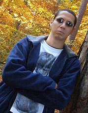
Premiado cientista na área de Psicologia Biológica, Onur Güntürkün afirma que o cérebro das aves pode ter design mais eficiente que o dos mamíferos (foto: Heiner Bayer)

26/05/2014

Por Karina Toledo

Agência FAPESP – Durante muito tempo predominou entre os neurocientistas a ideia de que o cérebro havia evoluído de forma linear. De acordo com a teoria proposta em meados do século 19 pelo neurologista alemão Ludwig Edinger (1855-1918), os peixes seriam os animais com o cérebro mais primitivo. Em seguida viriam os anfíbios, as aves e, finalmente, os mamíferos.

O cérebro dos mamíferos, segundo a teoria de Edinger, não apenas continha todas as estruturas existentes nos cérebros precedentes na escala evolutiva como também apresentava uma novidade que lhe proporcionava uma capacidade cognitiva superior e inédita: o neocórtex.

Mais desenvolvido nos primatas, o neocórtex é uma espécie de capa que recobre a parte externa do cérebro. Nos seres humanos, ele é dividido em seis camadas e apresenta uma grande quantidade de sulcos repletos de neurônios que comandam funções complexas como percepção sensorial, coordenação motora, raciocínio espacial e linguagem.

Para Edinger, como os pássaros não são dotados de neocórtex, jamais poderiam ser treinados como cachorros e gatos nem desenvolver habilidades cognitivas complexas, como usar ferramentas. Mas, no início do século 21, um grupo de cientistas demonstrou que essa teoria estava errada em artigo publicado no The Journal of Comparative Neurology.

Entre os autores estava Onur Güntürkün, professor da Ruhr-Universität Bochum, na Alemanha. Em outrapesquisa divulgada na revista PLoS Biology, Güntürkün mostrou que as gralhas são capazes de se reconhecer no espelho – algo que a maioria dos mamíferos não consegue fazer e que requer um certo grau de autoconsciência.

Por seu pioneirismo na área de Psicologia Biológica, Güntürkün, nascido na Turquia, recebeu em 2013 o Prêmio Gottfried Wilheim Leibniz, considerado o Nobel alemão. Em 2014, foi o ganhador doCommunicator Award, oferecido anualmente pela Deutsche Forschungsgemeinschaft (DFG) e pela Stifterverband für die Deutsche Wissenschaft a cientistas com boa habilidade de comunicar os resultados de sua pesquisa para um público amplo, fora da esfera científica.

No dia 20 de maio, Güntürkün esteve na sede da FAPESP para apresentar a palestra “Cognition without Cortex: The convergent evolution of avian and mammalian forebrains”, na qual contou que, a partir de seus experimentos com as gralhas, foi possível concluir que as aves teriam uma estrutura cerebral comparável ao neocórtex dos mamíferos.

“As aves possuem uma estrutura cerebral com as mesmas especificidades do neocórtex, a mesma bioquímica e o mesmo padrão de comunicação. A diferença é que não é dividida em camadas”, disse.

Segundo Güntürkün, é como se a natureza tivesse criado duas soluções diferentes para o mesmo problema (capacidade cognitiva avançada), em eventos distintos e independentes da história evolutiva.

“É possível que o design do cérebro das aves seja até mais eficiente do que o dos mamíferos, pois permite habilidades cognitivas complexas mesmo com um volume muito menor. No entanto, o cérebro das aves é pequeno demais para competir com o nosso”, avaliou.

Em entrevista concedida à Agência FAPESP, Güntürkün contou mais detalhes sobre suas pesquisas voltadas a entender as origens e a evolução do pensamento. Falou ainda sobre a importância da comunicação científica e sobre seus estudos relacionados às diferenças de gênero no cérebro.

Agência FAPESP – Qual é o motivo de sua visita ao Brasil?
Onur Güntürkün – Vim a convite da DFG para apresentar a “Palestra Leibniz” [formato desenvolvido para titulares do prêmio com o intuito de estimular o diálogo tanto com as comunidades científicas no exterior quanto com a sociedade em geral] e conversar com colegas cientistas do Brasil. O Brasil é um país muito importante, não apenas em termos de economia e política, mas também em termos de ciência. A Alemanha tem uma longa tradição em ciência, mas precisa planejar seu futuro de forma apropriada e, para isso, precisa pensar quais serão as grandes nações na área da ciência no futuro e como fomentar o relacionamento entre cientistas alemães e internacionais. Penso que o conceito da DFG é muito sábio: não são os diretores ou ministros que devem ser os embaixadores da ciência, mas os próprios cientistas. A única forma de isso ocorrer é possibilitando a interação entre eles, para que descubram interesses em comum. Dessa forma, é possível descobrir que, do outro lado do Atlântico, há uma ótima pessoa interessada nos mesmos assuntos que você e com quem você pode cooperar. Essa é a ideia.

Agência FAPESP – Como surgiu seu interesse pela evolução do cérebro e do pensamento?
Güntürkün – Só conseguimos entender alguma coisa quando conhecemos sua história. Só posso entender a mim mesmo quando sei algo sobre meu passado. O mesmo vale para o cérebro e a cognição. Se entendermos em quais condições evolutivas surgiram a cognição e o pensamento, podemos entender por que pensamos o que pensamos. Esta é a razão básica. Não me recordo de um momento de minha vida em que não estava interessado nesse assunto, então não há um ponto zero. Quando eu era criança já fazia ciência, realizava experimentos. Claro que eram simples e errados, sem conhecimento da literatura. Mas era ciência e foi um momento decisivo da minha existência. Muito do que faço hoje também deve estar errado e eu ainda não tenho consciência disso.

Agência FAPESP – Como forma e função estão relacionadas no cérebro? Até que ponto a estrutura cerebral define a capacidade de cognição?
Güntürkün – Se a arquitetura de nosso cérebro fosse diferente, nossa cognição seria diferente? A resposta é sim e não. Se perdêssemos um pedaço de nosso cérebro e nossa arquitetura fosse alterada, nossa cognição mudaria de forma radical. Há, no entanto, diferentes tipos de cérebros, com arquiteturas completamente diferentes, possivelmente capazes de criar o mesmo tipo de cognição. É como se você estivesse dirigindo um carro e perdesse uma parte do motor e ele deixasse de funcionar. Mas há outros tipos de motores que podem impulsionar um carro. Há diferentes soluções para o mesmo problema. Por isso, quando me perguntam se a estrutura define a cognição, minha resposta é sim e não. Sim – há diferentes soluções – e não – dentro de uma solução específica, todos os componentes precisam estar lá para o sistema funcionar. Essa é uma questão profunda de neurociência cognitiva. Podemos entender a evolução da cognição usando esse conhecimento como pano de fundo. Há diferentes organismos e diferentes tipos de cérebro. Eles pensam como nós ou possuem formas completamente diferentes de pensar que ainda não conhecemos? Isso é material suficiente para uma vida inteira de pesquisa.

Agência FAPESP – Em sua palestra, o senhor disse que as aves têm capacidades cognitivas comparáveis às dos mamíferos, embora não possuam o neocórtex. Isso ocorre com todas as aves ou apenas um grupo especial? E como isso é possível? 
Güntürkün – Acredito que nem todas as aves conseguem fazer isso, apenas algumas, como as gralhas e os corvos. E não sabemos por que as outras não têm essa habilidade. Mas o mesmo ocorre com os mamíferos. Os cachorros não se reconhecem no espelho, nem os gatos e nem mesmo os macacos rhesus. Apenas alguns mamíferos e alguns pássaros são capazes disso e ainda não sabemos ao certo a razão. O que há de especial no cérebro da gralha que o difere do cérebro do pombo? O que há de especial no cérebro do chimpanzé que lhe dá a capacidade de se reconhecer no espelho que o rhesus não tem? Não sabemos ainda. É uma questão profunda, pois, se o autorreconhecimento é uma pista para a consciência, poderemos entender melhor a consciência se formos capazes de entender como essas diferenças entre os animais aparecem.

Agência FAPESP – Teriam as aves uma espécie de neocórtex primitivo? 
Güntürkün – Não é primitivo. É como se a natureza tivesse inventado a roda duas vezes, de forma independente uma da outra. No cérebro das aves há uma estrutura interior virtualmente idêntica ao córtex pré-frontal humano. No entanto, ela não é dividida em camadas como o nosso córtex. Me parece que, em duas situações distintas na evolução, um grupo de animais precisou desenvolver altas capacidades cognitivas e terminou com o mesmo tipo de solução básica para esse problema. Mas um grupo desenvolveu o neocórtex e, o outro, um tipo diferente de estrutura cerebral. As invenções, porém, são absolutamente idênticas. É como ir a Marte e descobrir espécies completamente diferentes, com uma origem completamente diferente, mas, ao analisar profundamente, descobrir que alguns aspectos do cérebro dessas criaturas são virtualmente idênticos ao seu. É uma grande descoberta, pois sugere que não há duas soluções para um grande problema. Sempre se acaba inventando o mesmo tipo de roda quando se deseja criar um carro.

Agência FAPESP – O senhor sugeriu que o design do cérebro das aves talvez seja até mais eficiente que o dos mamíferos. Por quê? 
Güntürkün – É possível. De outra forma seria difícil entender como pequenos cérebros conseguem ser tão poderosos em termos cognitivos como o cérebro grande dos mamíferos. No entanto, o cérebro das aves nunca conseguiu ficar tão grande como o nosso. Não existe um único pássaro ou réptil que tenha sido capaz de desenvolver um cérebro de vários quilos. Não há nem sequer um réptil cujo cérebro pese mais do que 100 gramas. Não sabemos o porquê. Desde há mais de 300 milhões de anos, répteis e aves tiveram a chance de desenvolver um cérebro grande e nunca conseguiram. O argentinossauro, descoberto na Argentina, foi provavelmente o maior ser vivo que já habitou o planeta. Era enorme e tinha o cérebro do mesmo tamanho que o de um pássaro. O cérebro desses animais é restrito em termos de tamanho absoluto, enquanto nosso cérebro com a arquitetura cortical pode ficar grande. Essa foi a vantagem evolutiva que tivemos. De outra forma, estaríamos na gaiola e seríamos os animais de estimação das aves.

Agência FAPESP – O fato de o neocórtex corresponder a 76% do volume cerebral humano pode ser a explicação para sermos os animais mais inteligentes?
Güntürkün – Sim. É possível que tenhamos apenas um cérebro de primata muito grande. Simplesmente possuímos maior número de neurônios no neocórtex que qualquer outro animal do planeta. Há animais com cérebros maiores, como algumas baleias, elefantes, mas eles têm número menor de neurônios. Possivelmente nossa superioridade tenha razões quantitativas. É como os computadores. Colocamos mais memória, melhoramos outras especificações e, de repente, a máquina fica mais rápida, mais poderosa e capaz de calcular mais coisas.

Agência FAPESP – Somos, então, apenas primatas com um cérebro grande?
Güntürkün – Sim. Sou orgulhoso por ser um primata.

Agência FAPESP – O que já se conhece sobre o neocórtex e suas funções?
Güntürkün – Sabemos muito sobre o neocórtex, é uma das neuroestruturas mais bem estudadas. Por outro lado, entendemos muito pouco o cérebro dos pássaros. Obviamente, como somos mamíferos, acreditamos por centenas de anos que apenas com o neocórtex seria possível ter capacidades cognitivas avançadas, então havia um grande interesse em estudar o neocórtex. Agora que descobrimos que pássaros são tão capazes quanto nós, temos que trabalhar fortemente para preencher essa falta de conhecimento sobre as estruturas cerebrais das aves.

Agência FAPESP – Estudar o cérebro é sempre um grande desafio, pois não se pode simplesmente tirar um pedaço de tecido e analisar sob o microscópio sem grande consequências. Quais metodologias o senhor usa? 
Güntürkün – Claro que em humanos não podemos fazer experimentos invasivos, mas podemos gravar um eletroencefalograma, colocar nossos voluntários em uma máquina de ressonância magnética funcional e avaliar a atividade cerebral. Quando os pacientes têm má sorte ou genes ruins que resultam em uma alteração da estrutura cerebral, há sempre uma alteração correspondente nas habilidades cognitivas que podemos estudar. E, obviamente, fazemos experimentos comportamentais e coisas desse tipo. Nos animais, como os pombos que tenho usado muito no meu laboratório, podemos, mediante autorização, fazer experimentos invasivos, como implantar pequenos eletrodos no cérebro para gravar a atividade.

Agência FAPESP – O senhor tem estudos relacionados ao beijo e à tendência de os casais virarem a cabeça para a direita quando estão se beijando. Por que estudou esse tema? 
Güntürkün – É preciso deixar claro que não estudei o beijo para entender o beijo e sim para compreender a assimetria do cérebro humano. Tudo começou com a descoberta de que as aves têm um cérebro assimetricamente organizado. Essa assimetria aparece mesmo antes de saírem do ovo, quando viram a cabeça para o lado direito. Isso proporciona maior estimulação de luz no olho direito do embrião, que fica voltado para a casca do ovo. Então descobri, pela literatura, que humanos também viram a cabeça, desde antes de nascer, na maioria das vezes para o lado direito. E continuamos apresentando essa tendência por vários meses após o parto. Tenho uma teoria maluca de que isso, de alguma forma, modula nossos circuitos cerebrais. Se sou um recém-nascido, olho quase sempre para a direita, vejo minha mão direita e começo a fazer alguma atividade com a mão direita. E faço menos atividades com a mão esquerda. Então o fato de ser destro poderia ter sido influenciado por minha tendência de olhar para a direita.

Agência FAPESP – O senhor conseguiu comprovar essa teoria? 
Güntürkün – Formulei essa teoria e meus colegas me disseram que era bobagem, pois os bebês param de olhar para a direita por volta de 3 ou 4 meses de idade. E a destreza manual se manifesta muitos anos depois. Há um intervalo de tempo entre os dois eventos. Mas eu não acreditava nisso e pensei que talvez o padrão de movimentação dos bebês seja apenas muito complexo para que vejamos com clareza a tendência de virar a cabeça para a direita. Se essa tendência realmente nunca desaparece, nós, adultos, também devemos manifestá-la de alguma forma. Certo dia, eu estava sentado no sofá de minha casa e, de repente, me ocorreu: o beijo. Durante o ato de beijar não posso ficar com a cabeça reta, é preciso virá-la para um dos lados. Decidi observar casais em aeroportos enquanto eles estão esperando seus amados. O experimento foi feito em grandes aeroportos internacionais, de três diferentes continentes, para reduzir a possibilidade de qualquer viés cultural. Descobri que humanos têm a tendência de virar a cabeça para a direita em proporção absolutamente idêntica entre adultos e recém-nascidos: dois terços. Essa tendência não muda durante toda a vida e possivelmente ela modula a destreza manual nos humanos.

Agência FAPESP – No caso das aves, de certa forma, há uma relação com a estimulação do olho direito pela luz. E com os humanos? 
Güntürkün – Isso não acontece porque nossa visão é frontal. Minha teoria é que viramos a cabeça na tentativa de visualizar os próprios membros. Mas ainda não descobrimos o que mais é afetado por esse padrão de virar a cabeça além da destreza manual. É apenas um dos aspectos da assimetria cerebral que estamos estudando atualmente em meu laboratório.

Agência FAPESP – É verdade que há um lado do cérebro que controla emoções e habilidades com a música e outro lado responsável por atividades mais relacionadas com a razão?
Güntürkün – Isso é folclore existente na área de Psicologia e Neurociência. Precisamos de todo o cérebro para tocar uma música ou para raciocinar. Há algumas especializações relevantes. Para a música, por exemplo, nossa habilidade de compreender o ritmo é mais dominante no hemisfério direito. Então há um aspecto da música mais relacionado ao lado direito do cérebro. Depois que o famoso compositor [Maurice] Ravel teve um derrame no hemisfério direito, embora ainda fosse capaz de ouvir música, ele não conseguia mais compreendê-la, pois perdeu a habilidade de computar o ritmo. O raciocínio, porém, é algo que requer todo o cérebro. Muitos desses folclores possuem algum fundo de verdade, mas nem todos os fatos envolvidos são verdadeiros.

Agência FAPESP – Também é folclore que os homens têm mais neurônios do que as mulheres? 
Güntürkün – Isso é verdade. Os homens têm entre 10% e 15% mais neurônios, mesmo se o cálculo for proporcional ao tamanho do corpo. Mas a diferença na prática, francamente, ainda não sabemos. A inteligência de homens e mulheres é possivelmente idêntica. Há alguns cientistas que defendem que o QI [coeficiente de inteligência] é um pouco mais elevado nos homens. Se isso for verdade, no entanto, o efeito prático seria pequeno. Há outros estudos que não foram capazes de mostrar qualquer diferença. Minha teoria é que homens e mulheres são idênticos em termos de inteligência. Assumo essa premissa porque a maior parte da literatura mostra que, se há uma diferença, ela é muito pequena e não é importante. No entanto, é possível que exista diferença em termos de conhecimento. Homens aparentemente podem guardar um número de fatos cerca de 10% a 15% maior. Pode ser que o córtex seja um grande armazém e, se você tem um armazém maior, pode guardar mais itens dentro dele. Esta é minha teoria preferida e estamos elaborando estudos para analisá-la.

Agência FAPESP – Mas por que os homens precisariam de um armazém maior?
Güntürkün – Não tenho ideia. Não faz sentido em termos evolutivos. A pressão evolutiva de seleção, no que se refere ao conhecimento, atua sobre homens e mulheres de maneira igual. Por que os homens precisariam ter mais neurônios? Realmente não sei. Vamos morrer com muitas questões a serem respondidas. Mas, pelo menos, eu gostaria de descobrir se, de fato, existe uma relação entre ter mais neurônios e conseguir armazenar mais conhecimento. Aí uma questão ainda mais profunda apareceria: por quê? Fico ansioso de pensar que nunca vou saber.

Agência FAPESP – O senhor ganhou o Communicator Award de 2014, o que demonstra seu interesse em comunicar os resultados de sua pesquisa também a um público leigo. Por que acredita que a comunicação científica é importante?
Güntürkün – Estou muito honrado por ter sido escolhido. De acordo com o júri, sou capaz de me comunicar muito bem com a mídia e com o público em geral. Penso que isso é algo que todos nós, cientistas, temos de fazer. Precisamos falar sobre nossas pesquisas com a mídia, o público leigo e com outros cientistas e estudantes. E temos de fazer isso de forma que todos possam entender. É algo que considero meu dever, pois sou financiado pelos impostos dos contribuintes. Esses impostos garantem o melhor emprego do planeta a um número muito pequeno de pessoas: os cientistas. Trabalhamos naquilo que nos interessa, com quem desejamos e com as técnicas que escolhemos. Somos livres e podemos brincar com nossas ideias e isso é algo absolutamente fantástico. Ao mesmo tempo, somos rodeados por alunos brilhantes e muitas pessoas interessantes de todas as partes do mundo. Em troca, os contribuintes têm o direito de saber o que você está fazendo. E quando falo com esses trabalhadores não devo usar palavras que dificultem o entendimento. É meu dever.

Agência FAPESP – De forma geral, os cientistas cumprem bem esse dever? Como melhorar? 
Güntürkün – Acredito que, de maneira geral, os cientistas estão cientes desse dever e fazem um bom trabalho. Mas há algumas limitações. Há um imenso interesse em ciência por parte do público. Na televisão, não há apenas esportes e telenovelas, mas também programas sobre novas descobertas, animais e muitos outros aspectos relacionados à ciência. Jornalistas me procuram com frequência e a muitos de meus colegas. Fazem isso, obviamente, porque o jornalismo científico desperta interesse nas pessoas. Mas há uma responsabilidade dupla. Aos cientistas cabe não ter vergonha de falar com a mídia e ser claro. E a mídia tem a responsabilidade de divulgar a ciência da forma como ela é realmente, e não divulgar apenas escândalos, invenções fantásticas e coisas desse tipo.

Agência FAPESP – O senhor já teve problemas com a mídia? 
Güntürkün – Eu aprendi muito sobre a interação com a mídia ao longo de minha vida e tive algumas experiências difíceis. A maioria das pessoas da mídia realmente tenta fazer um bom trabalho. Mas, às vezes, os mecanismos internos da imprensa fazem com que as mensagens sejam muito simplificadas. Acho que esse é um problema que tanto cientistas quanto jornalistas precisam tentar solucionar de alguma forma.

New research helps explain how social understanding is performed by the brain (Science Daily)

Date:

February 24, 2014

Source: Aarhus University

Summary: An important question has been answered about how social understanding is performed in the brain. The findings may help us to attain a better understanding of why people with autism and schizophrenia have difficulties with social interaction. Using magnetic stimulation to temporarily disrupt normal processing of the areas of the human brain involved in the production of actions of human participants, it is demonstrated that these areas are also involved in the understanding of actions. The study is the first to demonstrate a clear causal effect, whereas earlier studies primarily have looked at correlations, which are difficult to interpret.

A new study from Aarhus University, Denmark, helps us understand why people with autism and schizophrenia have difficulties with social interaction. Credit: © styleuneed / Fotolia

In a study to be published in Psychological Science, researchers from Aarhus University and the University of Copenhagen demonstrate that brain cells in what is called the mirror system help people make sense of the actions they see other people perform in everyday life.

Using magnetic stimulation to temporarily disrupt normal processing of the areas of the human brain involved in the production of actions of human participants, it is demonstrated that these areas are also involved in the understanding of actions. The study is the first to demonstrate a clear causal effect, whereas earlier studies primarily have looked at correlations, which are difficult to interpret.

One of the researchers, John Michael, explains the process: “There has been a great deal of hype about the mirror system, and now we have performed an experiment that finally provides clear and straightforward evidence that the mirror system serves to help people make sense of others’ actions,” says John Michael.

Understanding autism and schizophrenia

The study shows that there are areas of the brain that are involved in the production of actions. And the researchers found evidence that these areas contribute to understanding others’ actions. This means that the same areas are involved in producing actions and understanding others’ actions. This helps us in everyday life, but it also holds great potential when trying to understand why people with autism and schizophrenia have difficulties with social interaction.

“Attaining knowledge of the processes underlying social understanding in people in general is an important part of the process of attaining knowledge of the underlying causes of the difficulties that some people diagnosed with autism and schizophrenia experience in sustaining social understanding. But it is important to emphasize that this is just one piece of the puzzle.”

“The findings may be interesting to therapists and psychiatrists who work with patients with schizophrenia or autism, or even to educational researchers,” adds John Michael.

Facts about the empirical basis

The participants (20 adults) came to the lab three times. They were given brain scans on the first visit. On the second and third, they received stimulation to their motor system and then performed a typical psychological task in which they watched brief videos of actors pantomiming actions (about 250 videos each time). After each video they had to choose a picture of an object that matched the pantomimed video. For example, a hammer was the correct answer for the video of an actor pretending to hammer.

This task was intended to gauge their understanding of the observed actions. The researchers found that the stimulation interfered with their performance of this task.

Innovative method

The researchers used an innovative technique for magnetically stimulating highly specific brain areas in order to temporarily disrupt normal processing in those areas. The reason for using this technique (called continuous theta-burst stimulation) in general is that it makes it possible to determine which brain areas perform which functions. For example, if you stimulate (and thus temporarily impair) area A, and the participants subsequently have difficulty with some specific task (task T), then you can infer that area A usually performs task T. The effect goes away after 20 minutes, so this is a harmless and widely applicable way to identify which tasks are performed by which areas.

With continuous theta-burst stimulation, you can actually determine that the activation of A contributes as a cause to people performing T. This method thus promises to be of great use to neuroscientists in the coming years.

Journal Reference:

  1. J. Michael, K. Sandberg, J. Skewes, T. Wolf, J. Blicher, M. Overgaard, C. D. Frith.Continuous Theta-Burst Stimulation Demonstrates a Causal Role of Premotor Homunculus in Action UnderstandingPsychological Science, 2014; DOI: 10.1177/0956797613520608

New ideas change your brain cells, research shows (Science Daily)

Date: 

February 24, 2014

Source: University of British Columbia

Summary: An important molecular change has been discovered that occurs in the brain when we learn and remember. The research shows that learning stimulates our brain cells in a manner that causes a small fatty acid to attach to delta-catenin, a protein in the brain. This biochemical modification is essential in producing the changes in brain cell connectivity associated with learning, the study finds. Findings may provide an explanation for some mental disabilities, the researchers say.

UBC’s Shernaz Bamji and Stefano Brigidi have discovered how brain cells change during learning and memories. Credit: UBC

A new University of British Columbia study identifies an important molecular change that occurs in the brain when we learn and remember.

Published this month in Nature Neuroscience, the research shows that learning stimulates our brain cells in a manner that causes a small fatty acid to attach to delta-catenin, a protein in the brain. This biochemical modification is essential in producing the changes in brain cell connectivity associated with learning, the study finds.

In animal models, the scientists found almost twice the amount of modified delta-catenin in the brain after learning about new environments. While delta-catenin has previously been linked to learning, this study is the first to describe the protein’s role in the molecular mechanism behind memory formation.

“More work is needed, but this discovery gives us a much better understanding of the tools our brains use to learn and remember, and provides insight into how these processes become disrupted in neurological diseases,” says co-author Shernaz Bamji, an associate professor in UBC’s Life Sciences Institute.

It may also provide an explanation for some mental disabilities, the researchers say. People born without the gene have a severe form of mental retardation called Cri-du-chat syndrome, a rare genetic disorder named for the high-pitched cat-like cry of affected infants. Disruption of the delta-catenin gene has also been observed in some patients with schizophrenia.

“Brain activity can change both the structure of this protein, as well as its function,” says Stefano Brigidi, first author of the article and a PhD candidate Bamji’s laboratory. “When we introduced a mutation that blocked the biochemical modification that occurs in healthy subjects, we abolished the structural changes in brain’s cells that are known to be important for memory formation.”

Journal Reference:

  1. G Stefano Brigidi, Yu Sun, Dayne Beccano-Kelly, Kimberley Pitman, Mahsan Mobasser, Stephanie L Borgland, Austen J Milnerwood, Shernaz X Bamji.Palmitoylation of δ-catenin by DHHC5 mediates activity-induced synapse plasticityNature Neuroscience, 2014; DOI: 10.1038/nn.3657

Cérebros humano e canino têm a mesma reação a vozes, sugere estudo (BBC)

Rebecca Morelle

Repórter de Ciências do BBC World Service

Atualizado em  22 de fevereiro, 2014 – 16:53 (Brasília) 19:53 GMT

Cachorros em aparelho de ressonância magnética (Borbala Ferenczy)

Estudo mostrou que a mesma região do cérebro de cães e humanos é ativada pelo som de vozes

Donos de cachorros costumam afirmar que seus animais de estimação conseguem entendê-los. Um novo estudo publicado no periódico Current Biology sugere que essas pessoas podem estar certas.

Ao colocar cães em um equipamento de ressonância magnética, pesquisadores húngaros descobriram que o cérebro desses animais reage da mesma forma que um cérebro humano a vozes de pessoas.

Outros sons carregados de emoção, como choro ou risadas, também geraram reações parecidas, o que talvez explica o fato de cachorros conseguirem se sintonizar às emoções de seus donos, afirmam os pesquisadores.

“Acreditamos que cães e humanos têm um mecanismo bastante similar para processar informações emocionais”, disse Attila Andics, da Universidade Eotvos Lorand e coordenador do estudo.

Sintonia

A pesquisa envolveu onze cães de estimação e comparou seus resultados aos de 22 voluntários humanos.

Para ambos os grupos, os cientistas tocaram 200 tipos diferentes sons, desde ruídos comuns, como o barulho de carros e de apitos, a sons emitidos por humanos (sem palavras) e por cães.

Cachorro em aparelho de ressonância magnética (Eniko Kubinyi)

Sons carregados de emoções, como risadas e choro, também geraram a mesma reação no cérebro dos cães e de pessoas

Os pesquisadores descobriram que uma região semelhante do cérebro – o polo temporal, que faz parte do lobo temporal – é ativada quando cães e pessoas ouvem vozes humanas.

“Já sabíamos que certas áreas no cérebro humano respondem mais fortemente a sons humanos do que a qualquer outro tipo de som”, explicou Andics. “É uma grande surpresa isso ocorrer também no cérebro canino. É a primeira vez que vemos algo assim em um animal que não seja um primata.”

O mesmo aconteceu quando sons como risadas e choros foram ouvidos. Uma área do cérebro conhecida como córtex auditivo primário foi ativada tanto em cachorros quanto em humanos.

Ao mesmo tempo, vocalizações caninas carregadas de emoção – como ganidos e latidos ferozes – também geraram uma reação parecida em todos os voluntários.

“Sabemos muito bem que cachorros conseguem se sintonizar ao sentimento de seus donos, e sabemos que um bom dono consegue identificar mudanças emocionais em seu cão – mas agora podemos começar a entender como isso é possível”, afirmou Andics.

No entanto, apesar dos cachorros reagirem à voz humana, suas reações foram bem mais fortes em relação aos sons caninos.

Os cães também parecem ser menos capazes de distinguir entre ruídos e sons vocais em comparação com humanos.

Palavras

Cães e aparelho de ressonância magnética (Eniko Kubinyi)

Próximo passo do estudo é checar como o cérebro de cães reage quando eles ouvem palavras

Ao comentar sobre a pesquisa, Sophie Scott, do Instituto de Neurociência Cognitiva da Universidade College London, disse: “Os cães são animais muito interessantes de se investigar porque muitos de seus traços desses os tornam dóceis em relação aos humanos. Alguns estudos mostram que eles entendem muitas palavras e o que queremos dizer quando apontamos para alguma coisa”.

Mas Scott acrescenta: “É algo bastante relevante encontrar isso em cães e não só em primatas, mas seria interessante também ver a reação desses animais a palavras. Risos e choros são parecidos com sons animais e por isso podem gerar esse tipo de reação.

“Um avanço seria demonstrar sensibilidade dos cães a palavras no idioma de seus donos.”

Segundo Andics, este será o foco da próxima série de testes da pesquisa.

Cientistas identificam gene que relaciona estrutura cerebral à inteligência (O Globo)

JC e-mail 4892, de 11 de fevereiro de 2014

Descoberta pode ter implicações importantes para a compreensão de transtornos psiquiátricos como esquizofrenia e autismo

Cientistas do King’s College London identificaram, pela primeira vez, um gene que relaciona a espessura da massa cinzenta do cérebro à inteligência. O estudo foi publicado nesta terça-feira na revista “Molecular Psychiatry” e pode ajudar a entender os mecanismos biológicos por trás de determinados danos intelectuais.

Até agora já se sabia que a massa cinzenta tinha um papel importante para a memória, atenção, pensamento, linguagem e consciência. Estudos anteriores também já mostravam que a espessura do córtex cerebral tinha a ver com a habilidade intelectual, mas nenhum gene tinha sido identificado.

Um time internacional de cientistas, liderado pelo King´s College, analisou amostras de DNA e exames de ressonância magnética por imagem de 1.583 adolescentes saudáveis de 14 anos, que também se submeteram a uma série de testes para determinar inteligência verbal e não verbal.

– Queríamos descobrir como diferenças estruturais no cérebro tinham a ver com diferenças na habilidade intelectual. Identificamos uma variação genética relacionada à plasticidade sináptica, de como os neurônios se comunicam – explica Sylvane Desrivières, principal autora do estudo, pelo Instituto de Psiquiatria do King’s College London. – Isto pode nos ajudar a entender o que acontece em nível neuronal com certas formas de comprometimento intelectual, onde a habilidade de comunicação dos neurônios é, de alguma forma, comprometida.

Ela acrescenta que é importante apontar que a inteligência é influenciada por muitos fatores genéticos e ambientais. O gene que identificamos só explica uma pequena proporção das diferenças nas habilidades intelectuais e não é, de forma alguma, “o gene da inteligência”.

Os pesquisadores observaram 54 mil possíveis variações envolvidas no desenvolvimento cerebral. Em média, adolescentes com uma variante genética particular tinham um córtex mais fino no hemisfério cerebral esquerdo, particularmente nos lobos frontal e temporal, e executavam bem testes de capacidade intelectual. A variação genética afeta a expressão do gene NPTN, que codifica uma proteína que atua nas sinapses neuronais e, portanto, afeta a forma como as células do cérebro se comunicam.

Para confirmar as suas conclusões, os pesquisadores estudaram o gene NPTN em células de camundongo e do cérebro humano. Os pesquisadores verificaram que o gene NPTN tinha uma atividade diferente nos hemisférios esquerdo e direito do cérebro, o que pode fazer com que o hemisfério esquerdo seja mais sensível aos efeitos das mutações NPTN. Os resultados sugerem que algumas diferenças na capacidade intelectual podem resultar da diminuição da função do gene NPTN em determinadas regiões do hemisfério esquerdo do cérebro.

A variação genética identificada neste estudo representa apenas uma estimativa de 0,5% da variação total em inteligência. No entanto, as descobertas podem ter implicações importantes para a compreensão dos mecanismos biológicos subjacentes de vários transtornos psiquiátricos, como esquizofrenia e autismo, nas quais a capacidade cognitiva é uma característica fundamental da doença.

http://oglobo.globo.com/ciencia/cientistas-identificam-gene-que-relaciona-estrutura-cerebral-inteligencia-11563313#ixzz2t1amCUSy

Brain regions thought to be uniquely human share many similarities with monkeys (Science Daily)

January 28, 2014

Source: Cell Press

Summary: New research suggests a surprising degree of similarity in the organization of regions of the brain that control language and complex thought processes in humans and monkeys. The study also revealed some key differences. The findings may provide valuable insights into the evolutionary processes that established our ties to other primates but also made us distinctly human.

 (A) The right vlFC ROI. Dorsally it included the inferior frontal sulcus and, more posteriorly, it included PMv; anteriorly it was bound by the paracingulate sulcus and ventrally by the lateral orbital sulcus and the border between the dorsal insula and the opercular cortex. (B) A schematic depiction of the result of the 12 cluster parcellation solution using an iterative parcellation approach. We subdivided PMv into ventral and dorsal regions (6v and 6r, purple and black). We delineated the IFJ area (blue) and areas 44d (gray) and 44v (red) in lateral pars opercularis. More anteriorly, we delineated areas 45 (orange) in the pars triangularis and adjacent operculum and IFS (green) in the inferior frontal sulcus and dorsal pars triangularis. We found area 12/47 in the pars orbitalis (light blue) and area Op (bright yellow) in the deep frontal operculum. We also identified area 46 (yellow), and lateral and medial frontal pole regions (FPl and FPm, ruby colored and pink). Credit: Neuron, Neubert et al.

New research suggests a surprising degree of similarity in the organization of regions of the brain that control language and complex thought processes in humans and monkeys. The study, publishing online January 28 in the Cell Press journal Neuron, also revealed some key differences. The findings may provide valuable insights into the evolutionary processes that established our ties to other primates but also made us distinctly human.

The research concerns the ventrolateral frontal cortex, a region of the brain known for more than 150 years to be important for cognitive processes including language, cognitive flexibility, and decision-making. “It has been argued that to develop these abilities, humans had to evolve a completely new neural apparatus; however others have suggested precursors to these specialized brain systems might have existed in other primates,” explains lead author Dr. Franz-Xaver Neubert of the University of Oxford, in the UK.

By using non-invasive MRI techniques in 25 people and 25 macaques, Dr. Neubert and his team compared ventrolateral frontal cortex connectivity and architecture in humans and monkeys. The investigators were surprised to find many similarities in the connectivity of these regions. This suggests that some uniquely human cognitive traits may rely on an evolutionarily conserved neural apparatus that initially supported different functions. Additional research may reveal how slight changes in connectivity accompanied or facilitated the development of distinctly human abilities.

The researchers also noted some key differences between monkeys and humans. For example, ventrolateral frontal cortex circuits in the two species differ in the way that they interact with brain areas involved with hearing.

“This could explain why monkeys perform very poorly in some auditory tasks and might suggest that we humans use auditory information in a different way when making decisions and selecting actions,” says Dr. Neubert.

A region in the human ventrolateral frontal cortex — called the lateral frontal pole — does not seem to have an equivalent area in the monkey. This area is involved with strategic planning, decision-making, and multi-tasking abilities.

“This might relate to humans being particularly proficient in tasks that require strategic planning and decision making as well as ‘multi-tasking’,” says Dr. Neubert.

Interestingly, some of the ventrolateral frontal cortex regions that were similar in humans and monkeys are thought to play roles in psychiatric disorders such as attention deficit hyperactivity disorder, obsessive compulsive disorder, and substance abuse. A better understanding of the networks that are altered in these disorders might lead to therapeutic insights.

Journal Reference:

  1. Franz-Xaver Neubert et al. Comparison of human ventral frontal cortex areas for cognitive control and language with areas in monkey frontal cortex.Neuron, Jan 28, 2014

Spirituality, Religion May Protect Against Major Depression by Thickening Brain Cortex (Science Daily)

Jan. 16, 2014 — A thickening of the brain cortex associated with regular meditation or other spiritual or religious practice could be the reason those activities guard against depression — particularly in people who are predisposed to the disease, according to new research led by Lisa Miller, professor and director of Clinical Psychology and director of the Spirituality Mind Body Institute at Teachers College, Columbia University.

The study, published online by JAMA Psychiatry, involved 103 adults at either high or low risk of depression, based on family history. The subjects were asked how highly they valued religion or spirituality. Brain MRIs showed thicker cortices in subjects who placed a high importance on religion or spirituality than those who did not. The relatively thicker cortex was found in precisely the same regions of the brain that had otherwise shown thinning in people at high risk for depression.

Although more research is necessary, the results suggest that spirituality or religion may protect against major depression by thickening the brain cortex and counteracting the cortical thinning that would normally occur with major depression. The study, published on Dec. 25, 2013, is the first published investigation on the neuro-correlates of the protective effect of spirituality and religion against depression.

“The new study links this extremely large protective benefit of spirituality or religion to previous studies which identified large expanses of cortical thinning in specific regions of the brain in adult offspring of families at high risk for major depression,” Miller said.

Previous studies by Miller and the team published in theAmerican Journal of Psychiatry (2012) showed a 90 percent decrease in major depression in adults who said they highly valued spirituality or religiosity and whose parents suffered from the disease. While regular attendance at church was not necessary, a strong personal importance placed on spirituality or religion was most protective against major depression in people who were at high familial risk.

Journal Reference:

  1. Lisa Miller, Ravi Bansal, Priya Wickramaratne, Xuejun Hao, Craig E. Tenke, Myrna M. Weissman, Bradley S. Peterson.Neuroanatomical Correlates of Religiosity and SpiritualityJAMA Psychiatry, 2013; : 1 DOI:10.1001/jamapsychiatry.2013.3067

Brain Regions ‘Tune’ Activity to Enable Attention (Science Daily)

Jan. 16, 2014 — The brain appears to synchronize the activity of different brain regions to make it possible for a person to pay attention or concentrate on a task, scientists at Washington University School of Medicine in St. Louis have learned.

 Scientists at the Neuroimaging Laboratory at Washington University School of Medicine in St. Louis have learned that brain regions sync their activity levels to enable attention. Pictured (from left) are first author Amy Daitch, co-senior author Maurizio Corbetta, postdoctoral researcher Alicia Callejas and McDonnell Scholar Lenny Ramsey. (Credit: Robert J. Boston)

Researchers think the process, roughly akin to tuning multiple walkie-talkies to the same frequency, may help establish clear channels for communication between brain areas that detect sensory stimuli.

“We think the brain not only puts regions that facilitate attention on alert but also makes sure those regions have open lines for calling each other,” said first author Amy Daitch, a graduate student researcher.

The results are available in the Proceedings of the National Academy of Sciences.

People who suffer from brain injuries or strokes often have problems paying attention and concentrating.

“Attention deficits in brain injury have been thought of as a loss of the resources needed to concentrate on a task,” said senior author Maurizio Corbetta, MD, the Norman J. Stupp Professor of Neurology. “However, this study shows that temporal alignment of responses in different brain areas is also a very important mechanism that contributes to attention and could be impaired by brain injury.”

Attention lets people ignore irrelevant sensory stimuli, like a driver disregarding a ringing cell phone, and pay attention to important stimuli, like a deer stepping onto the road in front of the car.

To analyze brain changes linked to attention, the scientists used grids of electrodes temporarily implanted onto the brains of patients with epilepsy. Co-senior author Eric Leuthardt, MD, associate professor of neurosurgery and bioengineering, uses the grids to map for surgical removal of brain tissues that contribute to uncontrollable seizures.

With patient permission, the grids also can allow Leuthardt’s lab to study human brain activity at a level of detail unavailable via any other method. Normally, Corbetta and his colleagues investigate attention using various forms of magnetic resonance imaging (MRI), which can detect changes in brain activity that occur every 2 to 3 seconds. But with the grids in place, Corbetta and Leuthardt can study the changes that occur in milliseconds.

Before grid implantation, the scientists scanned the brains of seven epilepsy patients, using MRI to map regions known to contribute to attention. With the grids in place, the researchers monitored brain cells as the patients watched for visual targets, directing their attention to different locations on a computer screen without moving their eyes. When patients saw the targets, they pressed a button to let the scientists know they had seen them.

“We analyzed brain oscillations that reflect fluctuations in excitability of a local brain region; in other words, how difficult or easy it is for a neuron to respond to an input,” Daitch said. “If areas of the brain involved in detecting a stimulus are at maximum excitability, you would be much more likely to notice the stimulus.”

Excitability regularly rises and falls in the cells that make up a given brain region. But these oscillations normally are not aligned between different brain regions.

The researchers’ results showed that as patients directed their attention, the brain regions most important for paying attention to visual stimuli adjusted their excitability cycles, causing them to start hitting the peaks of their cycles at the same time. In regions not involved in attention, the excitability cycles did not change.

“If the cycles of two brain regions are out of alignment, the chances that a signal from one region will get through to another region are reduced,” Corbetta said.

Daitch, Corbetta and Leuthardt are investigating whether knowing not just the location, but also the tempo of the task, allows participants to bring the excitability of their brain regions into alignment more rapidly.

Journal Reference:

  1. A. L. Daitch, M. Sharma, J. L. Roland, S. V. Astafiev, D. T. Bundy, C. M. Gaona, A. Z. Snyder, G. L. Shulman, E. C. Leuthardt, M. Corbetta. Frequency-specific mechanism links human brain networks for spatial attention.Proceedings of the National Academy of Sciences, 2013; 110 (48): 19585 DOI: 10.1073/pnas.1307947110

Discovery of Quantum Vibrations in ‘Microtubules’ Inside Brain Neurons Supports Controversial Theory of Consciousness (Science Daily)

Jan. 16, 2014 — A review and update of a controversial 20-year-old theory of consciousness published in Physics of Life Reviews claims that consciousness derives from deeper level, finer scale activities inside brain neurons. The recent discovery of quantum vibrations in “microtubules” inside brain neurons corroborates this theory, according to review authors Stuart Hameroff and Sir Roger Penrose. They suggest that EEG rhythms (brain waves) also derive from deeper level microtubule vibrations, and that from a practical standpoint, treating brain microtubule vibrations could benefit a host of mental, neurological, and cognitive conditions.

A review and update of a controversial 20-year-old theory of consciousness published in Physics of Life Reviews claims that consciousness derives from deeper level, finer scale activities inside brain neurons. (Credit: © James Steidl / Fotolia)

The theory, called “orchestrated objective reduction” (‘Orch OR’), was first put forward in the mid-1990s by eminent mathematical physicist Sir Roger Penrose, FRS, Mathematical Institute and Wadham College, University of Oxford, and prominent anesthesiologist Stuart Hameroff, MD, Anesthesiology, Psychology and Center for Consciousness Studies, The University of Arizona, Tucson. They suggested that quantum vibrational computations in microtubules were “orchestrated” (“Orch”) by synaptic inputs and memory stored in microtubules, and terminated by Penrose “objective reduction” (‘OR’), hence “Orch OR.” Microtubules are major components of the cell structural skeleton.

Orch OR was harshly criticized from its inception, as the brain was considered too “warm, wet, and noisy” for seemingly delicate quantum processes.. However, evidence has now shown warm quantum coherence in plant photosynthesis, bird brain navigation, our sense of smell, and brain microtubules. The recent discovery of warm temperature quantum vibrations in microtubules inside brain neurons by the research group led by Anirban Bandyopadhyay, PhD, at the National Institute of Material Sciences in Tsukuba, Japan (and now at MIT), corroborates the pair’s theory and suggests that EEG rhythms also derive from deeper level microtubule vibrations. In addition, work from the laboratory of Roderick G. Eckenhoff, MD, at the University of Pennsylvania, suggests that anesthesia, which selectively erases consciousness while sparing non-conscious brain activities, acts via microtubules in brain neurons.

“The origin of consciousness reflects our place in the universe, the nature of our existence. Did consciousness evolve from complex computations among brain neurons, as most scientists assert? Or has consciousness, in some sense, been here all along, as spiritual approaches maintain?” ask Hameroff and Penrose in the current review. “This opens a potential Pandora’s Box, but our theory accommodates both these views, suggesting consciousness derives from quantum vibrations in microtubules, protein polymers inside brain neurons, which both govern neuronal and synaptic function, and connect brain processes to self-organizing processes in the fine scale, ‘proto-conscious’ quantum structure of reality.”

After 20 years of skeptical criticism, “the evidence now clearly supports Orch OR,” continue Hameroff and Penrose. “Our new paper updates the evidence, clarifies Orch OR quantum bits, or “qubits,” as helical pathways in microtubule lattices, rebuts critics, and reviews 20 testable predictions of Orch OR published in 1998 — of these, six are confirmed and none refuted.”

An important new facet of the theory is introduced. Microtubule quantum vibrations (e.g. in megahertz) appear to interfere and produce much slower EEG “beat frequencies.” Despite a century of clinical use, the underlying origins of EEG rhythms have remained a mystery. Clinical trials of brief brain stimulation aimed at microtubule resonances with megahertz mechanical vibrations using transcranial ultrasound have shown reported improvements in mood, and may prove useful against Alzheimer’s disease and brain injury in the future.

Lead author Stuart Hameroff concludes, “Orch OR is the most rigorous, comprehensive and successfully-tested theory of consciousness ever put forth. From a practical standpoint, treating brain microtubule vibrations could benefit a host of mental, neurological, and cognitive conditions.”

The review is accompanied by eight commentaries from outside authorities, including an Australian group of Orch OR arch-skeptics. To all, Hameroff and Penrose respond robustly.

Penrose, Hameroff and Bandyopadhyay will explore their theories during a session on “Microtubules and the Big Consciousness Debate” at the Brainstorm Sessions, a public three-day event at the Brakke Grond in Amsterdam, the Netherlands, January 16-18, 2014. They will engage skeptics in a debate on the nature of consciousness, and Bandyopadhyay and his team will couple microtubule vibrations from active neurons to play Indian musical instruments. “Consciousness depends on anharmonic vibrations of microtubules inside neurons, similar to certain kinds of Indian music, but unlike Western music which is harmonic,” Hameroff explains.

Journal References:

  1. Stuart Hameroff and Roger Penrose. Consciousness in the universe: A review of the ‘Orch OR’ theoryPhysics of Life Reviews, 2013 DOI: 10.1016/j.plrev.2013.08.002
  2. Stuart Hameroff, MD, and Roger Penrose. Reply to criticism of the ‘Orch OR qubit’–‘Orchestrated objective reduction’ is scientifically justifiedPhysics of Life Reviews, 2013 DOI: 10.1016/j.plrev.2013.11.00
  3. Stuart Hameroff, Roger Penrose. Consciousness in the universePhysics of Life Reviews, 2013; DOI:10.1016/j.plrev.2013.08.002

SHY Hypothesis Explains That Sleep Is the Price We Pay for Learning (Science Daily)

Jan. 9, 2014 — Why do animals ranging from fruit flies to humans all need to sleep? After all, sleep disconnects them from their environment, puts them at risk and keeps them from seeking food or mates for large parts of the day.

Sleeping puppy. Is sleep the price the brain must pay for learning and memory? (Credit: © paul prescott / Fotolia)

Two leading sleep scientists from the University of Wisconsin School of Medicine and Public Health say that their synaptic homeostasis hypothesis of sleep or “SHY” challenges the theory that sleep strengthens brain connections. The SHY hypothesis, which takes into account years of evidence from human and animal studies, says that sleep is important because it weakens the connections among brain cells to save energy, avoid cellular stress, and maintain the ability of neurons to respond selectively to stimuli.

“Sleep is the price the brain must pay for learning and memory,” says Dr. Giulio Tononi, of the UW Center for Sleep and Consciousness. “During wake, learning strengthens the synaptic connections throughout the brain, increasing the need for energy and saturating the brain with new information. Sleep allows the brain to reset, helping integrate, newly learned material with consolidated memories, so the brain can begin anew the next day. ”

Tononi and his co-author Dr. Chiara Cirelli, both professors of psychiatry, explain their hypothesis in a review article in today’s issue of the journal Neuron. Their laboratory studies sleep and consciousness in animals ranging from fruit flies to humans; SHY takes into account evidence from molecular, electrophysiological and behavioral studies, as well as from computer simulations. “Synaptic homeostasis” refers to the brain’s ability to maintain a balance in the strength of connections within its nerve cells.

Why would the brain need to reset? Suppose someone spent the waking hours learning a new skill, such as riding a bike. The circuits involved in learning would be greatly strengthened, but the next day the brain will need to pay attention to learning a new task. Thus, those bike-riding circuits would need to be damped down so they don’t interfere with the new day’s learning.

“Sleep helps the brain renormalize synaptic strength based on a comprehensive sampling of its overall knowledge of the environment,” Tononi says, “rather than being biased by the particular inputs of a particular waking day.”

The reason we don’t also forget how to ride a bike after a night’s sleep is because those active circuits are damped down less than those that weren’t actively involved in learning. Indeed, there is evidence that sleep enhances important features of memory, including acquisition, consolidation, gist extraction, integration and “smart forgetting,” which allows the brain to rid itself of the inevitable accumulation of unimportant details. However, one common belief is that sleep helps memory by further strengthening the neural circuits during learning while awake. But Tononi and Cirelli believe that consolidation and integration of memories, as well as the restoration of the ability to learn, all come from the ability of sleep to decrease synaptic strength and enhance signal-to-noise ratios.

While the review finds testable evidence for the SHY hypothesis, it also points to open issues. One question is whether the brain could achieve synaptic homeostasis during wake, by having only some circuits engaged, and the rest off-line and thus resetting themselves. Other areas for future research include the specific function of REM sleep (when most dreaming occurs) and the possibly crucial role of sleep during development, a time of intense learning and massive remodeling of brain.

This work was supported by NIMH (1R01MH091326 and 1R01MH099231 to GT and CC)

Journal Reference:

  1. Giulio Tononi, Chiara Cirelli. Sleep and the Price of Plasticity: From Synaptic and Cellular Homeostasis to Memory Consolidation and IntegrationNeuron, 2014; 81 (1): 12 DOI: 10.1016/j.neuron.2013.12.025

A mulher que encolheu o cérebro humano (O Globo)

Suzana Herculano é a primeira brasileira a falar na prestigiada conferência TED

Ela debaterá o cérebro de 86 bilhões de neurônios (e não 100 bilhões, como se acreditava) e como o homem se diferenciou dos primatas 

Publicado:24/05/13 – 7h00; Atualizado:24/05/13 – 11h41

Suzana Herculano-Houzel, professora do Instituto de Ciências Biomédicas da UFRJFoto: Guito Moreto

Suzana Herculano-Houzel, professora do Instituto de Ciências Biomédicas da UFRJ Guito Moreto

Neurocientista da UFRJ, Suzana Herculano-Houzel é a primeira brasileira a participar da TED (Tecnologia, Entretenimento e Design, em português) — prestigiada série de conferências que reúne grandes nomes das mais diversas áreas do conhecimento para debater novas ideias. Suzana falará no dia 12 de junho, sob o tema “Ouça a natureza”, e destacará suas descobertas únicas sobre o cérebro humano.

Sobre o que vai falar na TED?

Vou falar sobre o cérebro humano e mostrar como ele não é um cérebro especial, uma exceção à regra. Nossas pesquisas nos revelaram que se trata apenas de um cérebro de primata grande. O notável é que passamos a ter um cérebro enorme, do tamanho que nenhum outro primata tem, nem os maiores, porque inventamos o cozimento dos alimentos e, com isso, passamos a ter um número enorme de neurônios.

O cozimento foi fundamental para nos tornarmos humanos?

Sim, burlamos a limitação energética imposta pela dieta crua. E a implicação bacana e irônica é que, com isso, conseguimos liberar tempo no cérebro para nos dedicarmos a outras coisas (que não buscar alimentos), como criar a agricultura, as civilizações, a geladeira e a eletricidade. Até o ponto em que conseguir comida cozida e calorias em excesso ficou tão fácil que, agora, temos o problema inverso: estamos comendo demais. Por isso, voltamos à saladinha.

Se alimentarmos orangotangos e gorilas com comida cozida eles serão tão inteligentes quanto nós?

Sim, porque não seriam limitados pelo número reduzido de calorias que conseguem com a comida crua. Claro que nós fizemos uma inovação cultural ao inventar a cozinha. Tem uma diferença entre dar comida cozida para o animal e ele ter o desenvolvimento cultural do cozimento. Mas, ainda assim, se em todas as refeições eles tiverem acesso à comida cozida, daqui a 200 mil ou 300 mil anos eles terão o cérebro maior. Com a alimentação que têm hoje, não é possível terem um cérebro maior dado o corpo grande que têm. É uma coisa ou outra.

Somos especiais?

A gente não é especial coisa alguma. Somos apenas um primata que burlou as regras energéticas e conseguiu botar mais neurônios no cérebro de um jeito que nenhum outro animal conseguiu. Por isso estudamos os outros animais e não o contrário.

Persistem ainda mitos sobre o cérebro? Como o dos 100 bilhões de neurônios, que seus estudos demonstraram que são, na verdade, 86 bilhões?

Sim, eles continuam existindo, mesmo na neurociência. O nosso trabalho já é muito citado como referência. As coisas estão mudando. E o mais legal é que é por conta da ciência tupiniquim, o que eu acho maravilhoso. Mas vemos que é um processo, que ainda tem muita gente que insiste no número antigo.

O novo manual de diagnóstico de doenças mentais dos EUA (que serve de referência para todo o mundo, inclusive para a OMS) foi lançado na semana passada em meio à controvérsia. Especialistas acham que são tantos transtornos que praticamente não resta mais nenhum espaço para a normalidade. Qual a sua opinião?

Acho que essa discussão é muito necessária, justamente para reconhecermos o que são as variações ao redor do normal e quais são os extremos problemáticos e doentios de fato. Então, a discussão é importante, ótima a qualquer momento. Mas acho também que há muita informação errada e sensacionalista circulando, sobretudo sobre o déficit de atenção. As estatísticas variam muito de país para país, às vezes porque varia o número de médicos que reconhece a criança como portadora do distúrbio. E acho que ainda há um problema enorme, um medo enorme do estereótipo da doença mental. Até hoje ainda existe uma resistência louca em ir a um psiquiatra. E acho que, pelo contrário, ganhamos muito reconhecendo que existem transtornos e que eles podem ser tratados.

Ainda há muito estigma?

O maior problema hoje em dia é que é feio ter um distúrbio no cérebro. Perceba que nem estou falando em transtorno mental. Precisar de remédio para o cérebro é terrível. E temos tanto a ganhar reconhecendo os problemas, fazendo os diagnósticos. O cérebro é tão complexo, tem tanta coisa para dar errado, que o espantoso é que não dê problema em todo mundo sempre. Então, acho normal que boa parte da população tenha algum problema, não me espanta nem um pouco. E, uma vez que se reconhece o problema, que se faz o diagnóstico, há a opção de poder tratar. Se dispomos de um tratamento, por que não usar?

O presidente dos EUA, Barack Obama, recentemente anunciou uma inédita iniciativa de reunir pesquisadores dos mais diversos centros para estudar exclusivamente o cérebro. O que podemos esperar de tamanho esforço científico?

Não só o cérebro, mas o cérebro em atividade. Obama quer ir além do que já tinham feito — estudar a função de diferentes áreas — e entender como se conectam, como falam umas com as outras, ter ideia desse funcionamento integrado, dessa interação. Essa é uma das grandes lacunas do conhecimento: entender como as várias partes do cérebro funcionam ao mesmo tempo. Não sabemos como o cérebro funciona como um todo; é uma das fronteiras finais do conhecimento.

Não sabemos como o cérebro funciona?

Como um todo, não. Sabemos o que as partes fazem, mas não sabemos como se dá a conversa entre elas. Não sabemos a origem da consciência, da sensação do “eu estou aqui agora”. Que áreas são fundamentais para isso? É esse tipo de conhecimento que se está buscando, do cérebro funcionando ao vivo e em cores, em tempo real.

O objetivo não é estudar doenças, então?

Não, o grande objetivo é estudar consciência, memória; entender como o cérebro reúne emoção e lógica, coisas que são fruto da ação coordenada de várias partes. Claro que desse conhecimento todo podem surgir implicações para o Alzheimer e outras doenças. Mas, na verdade, falar em doenças é uma roupagem usada pela divulgação do programa para o público assimilar melhor. Existe esse preconceito de que a ciência só vale quando resolve uma doença.

Leia mais sobre esse assunto em http://oglobo.globo.com/ciencia/a-mulher-que-encolheu-cerebro-humano-8482825#ixzz2UFWUvdYn © 1996 – 2013. Todos direitos reservados a Infoglobo Comunicação e Participações S.A. Este material não pode ser publicado, transmitido por broadcast, reescrito ou redistribuído sem autorização.

Schizophrenia Symptoms Eliminated in Animal Model (Science Daily)

May 22, 2013 — Overexpression of a gene associated with schizophrenia causes classic symptoms of the disorder that are reversed when gene expression returns to normal, scientists report. 

Overexpression of a gene associated with schizophrenia causes classic symptoms of the disorder that are reversed when gene expression returns to normal, scientists report. Pictured are (left to right) Drs. Lin Mei, Dongmin Yin and Yongjun Chen, Medical College of Georgia at Georgia Regents University. (Credit: Phil Jones, Georgia Regents University Photographer)

They genetically engineered mice so they could turn up levels of neuregulin-1 to mimic high levels found in some patients then return levels to normal, said Dr. Lin Mei, Director of the Institute of Molecular Medicine and Genetics at the Medical College of Georgia at Georgia Regents University.

They found that when elevated, mice were hyperactive, couldn’t remember what they had just learned and couldn’t ignore distracting background or white noise. When they returned neuregulin-1 levels to normal in adult mice, the schizophrenia-like symptoms went away, said Mei, corresponding author of the study in the journal Neuron.

While schizophrenia is generally considered a developmental disease that surfaces in early adulthood, Mei and his colleagues found that even when they kept neuregulin-1 levels normal until adulthood, mice still exhibited schizophrenia-like symptoms once higher levels were expressed. Without intervention, they developed symptoms at about the same age humans do.

“This shows that high levels of neuregulin-1 are a cause of schizophrenia, at least in mice, because when you turn them down, the behavior deficit disappears,” Mei said. “Our data certainly suggests that we can treat this cause by bringing down excessive levels of neuregulin-1 or blocking its pathologic effects.”

Schizophrenia is a spectrum disorder with multiple causes — most of which are unknown — that tends to run in families, and high neuregulin-1 levels have been found in only a minority of patients. To reduce neuregulin-1 levels in those individuals likely would require development of small molecules that could, for example, block the gene’s signaling pathways, Mei said. Current therapies treat symptoms and generally focus on reducing the activity of two neurotransmitters since the bottom line is excessive communication between neurons.

The good news is it’s relatively easy to measure neuregulin-1 since blood levels appear to correlate well with brain levels. To genetically alter the mice, they put a copy of the neuregulin-1 gene into mouse DNA then, to make sure they could control the levels, they put in front of the DNA a binding protein for doxycycline, a stable analogue for the antibiotic tetracycline, which is infamous for staining the teeth of fetuses and babies.

The mice are born expressing high levels of neuregulin-1 and giving the antibiotic restores normal levels. “If you don’t feed the mice tetracycline, the neuregulin-1 levels are always high,” said Mei, noting that endogenous levels of the gene are not affected. High-levels of neuregulin-1 appear to activate the kinase LIMK1, impairing release of the neurotransmitter glutamate and normal behavior. The LIMK1 connection identifies another target for intervention, Mei said.

Neuregulin-1 is essential for heart development as well as formation of myelin, the insulation around nerves. It’s among about 100 schizophrenia-associated genes identified through genome-wide association studies and has remained a consistent susceptibility gene using numerous other methods for examining the genetics of the disease. It’s also implicated in cancer.

Mei and his colleagues were the first to show neuregulin-1’s positive impact in the developed brain, reporting in Neuron in 2007 that it and its receptor ErbB4 help maintain a healthy balance of excitement and inhibition by releasing GABA, a major inhibitory neurotransmitter, at the sight of inhibitory synapses, the communication paths between neurons. Years before, they showed the genes were also at excitatory synapses, where they also could quash activation. In 2009, the MCG researchers provided additional evidence of the role of neuregulin-1 in schizophrenia by selectively deleting the gene for its receptor, ErbB4 and creating another symptomatic mouse.

Schizophrenia affects about 1 percent of the population, causing hallucinations, depression and impaired thinking and social behavior. Babies born to mothers who develop a severe infection, such as influenza or pneumonia, during pregnancy have a significantly increased risk of schizophrenia.

Journal Reference:

  1. Dong-Min Yin, Yong-Jun Chen, Yi-Sheng Lu, Jonathan C. Bean, Anupama Sathyamurthy, Chengyong Shen, Xihui Liu, Thiri W. Lin, Clifford A. Smith, Wen-Cheng Xiong, Lin Mei.Reversal of Behavioral Deficits and Synaptic Dysfunction in Mice Overexpressing Neuregulin 1.Neuron, 2013; 78 (4): 644 DOI:10.1016/j.neuron.2013.03.028